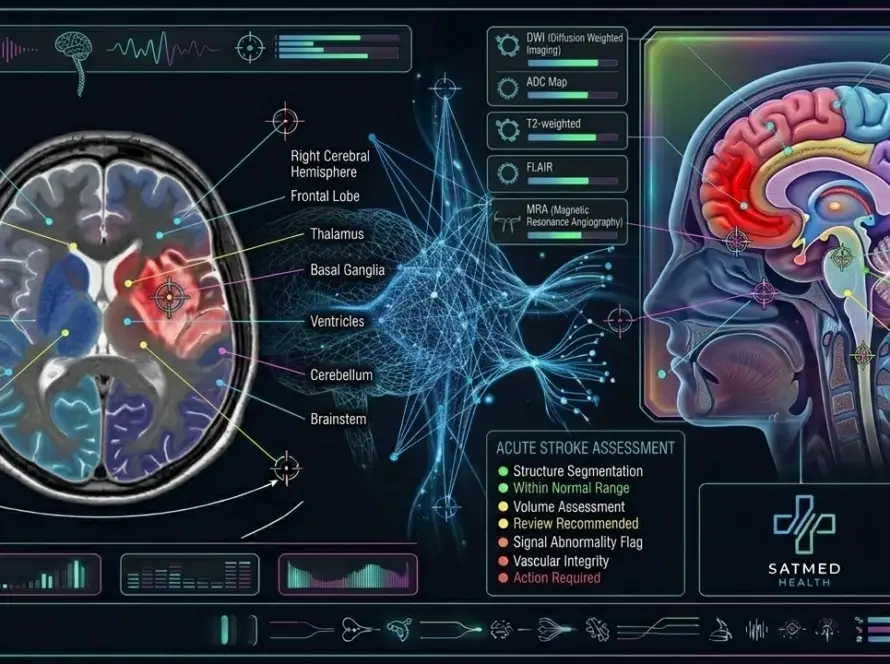
Why remaining completely still and holding your breath when instructed is vital to prevent motion blur and ensure high-resolution diagnostic images.
Positioning & Breath-Hold in Radiology: Why Staying Still Matters
At a glance
- Even tiny movements during a scan can blur images and hide subtle abnormalities, just like a shaky camera photo.
- Breath-holds of 5–15 seconds freeze diaphragm, liver, and lung movement for crystal-clear images.
- Positioning aids like foam pads, knee wedges, and head restraints improve comfort and reduce the urge to shift.
- If you accidentally move or cough, technologists simply re-run the affected sequence—no need to restart the entire exam.
- Each imaging modality—CT, MRI, X-ray, and ultrasound—has unique positioning requirements tailored to the body part being studied.
Why do I need to stay still during a CT scan? This is one of the most common questions patients ask when they arrive at the imaging center. The answer is simple but critical: motion is the enemy of clarity. Whether you are having an X-ray, CT, MRI, or ultrasound, even the tiniest shift in position can turn a potentially diagnostic image into a blurry mess that forces the entire sequence to be repeated.
Clinical context: The American College of Radiology (ACR) and the Society of Radiographers identify patient motion as one of the top three causes of non-diagnostic imaging studies. Proper positioning and breath-hold compliance directly impact image quality, diagnostic confidence, and patient safety.
This guide explains the science behind radiology positioning and breath-hold techniques, what technologists do to keep you comfortable and motionless, and exactly what to do if you accidentally cough, sneeze, or shift during your scan. Understanding these principles transforms an anxious experience into a collaborative effort between you and your imaging team.
🚀 Elevate Your Department's Imaging Standards
Access premium interventional radiology consumables and safety solutions designed for modern cath labs and imaging suites.
Explore SATMED Health Solutions →Preventing image motion blur
Think of a medical scanner like a high-speed camera. Just as a photograph of a moving car becomes a streaky blur, any movement during an imaging acquisition creates motion artifacts—streaks, ghosting, and loss of fine detail that can obscure the very abnormalities your doctor is looking for.
How motion affects different modalities
- CT scans: The X-ray tube rotates around you in less than one second per slice. Even a slight wiggle during this rotation creates double images and streak artifacts across the entire slice.
- MRI: Each image sequence can take 2–5 minutes to acquire. Because MRI builds images from thousands of individual signal measurements, any movement during that window causes ghosting artifacts that propagate across the entire image.
- X-rays: A standard chest or bone X-ray captures in a fraction of a second, so brief movement is less problematic. However, poor positioning—such as a rotated shoulder—can distort anatomical relationships and lead to misdiagnosis.
- Ultrasound: Real-time imaging means motion is expected, but the sonographer needs you to hold specific positions steady so they can capture diagnostic still frames and measurements.
Consequence of motion: When an image is degraded by patient movement, the radiologist may not be able to answer the clinical question. This can lead to a repeat scan, delayed diagnosis, or in some cases, a more invasive procedure that could have been avoided with a clear initial image.
💉 Optimise Your Cath Lab Workflow
SATLine consumables deliver consistent performance across diagnostic and interventional procedures. Reduce waste and standardise inventory.
Discover SATLine Products →Why radiologists ask you to hold your breath
Breathing is something we do roughly 20,000 times per day without thinking. But during a diagnostic scan, each breath moves your diaphragm, shifts your liver, expands your lungs, and displaces abdominal organs by several centimeters. For the radiologist, that movement is the difference between a crisp, diagnostic image and a useless blur.
What happens when you breathe during a scan
When you inhale, your diaphragm drops and your lungs expand, pushing the liver and spleen downward. When you exhale, everything returns upward. Over a 5-second scan, this up-and-down motion creates a smeared, ghost-like duplicate of every organ on the image. A breath-hold freezes everything in place, allowing the scanner to capture a single, sharp snapshot.
Typical breath-hold duration by scan type
- Chest CT: 5–10 seconds
- Abdomen/pelvis CT: 10–15 seconds
- Cardiac CT angiography: 5–8 seconds (often with automated breath-hold coaching)
- MRI (most sequences): 10–20 seconds per breath-hold interval
- X-ray: Usually no breath-hold needed; image capture is instantaneous
Diagnostic pearl: Modern CT scanners are incredibly fast. A full chest scan can be completed in a single 5-second breath-hold, and some advanced machines can image the entire heart in just one heartbeat. The faster the scanner, the shorter your breath-hold needs to be.
Positioning aids and comfort devices
Imaging technologists use a variety of positioning aids to keep you comfortable, properly aligned, and motionless throughout your exam. These devices are not optional extras—they are essential tools that improve image quality and reduce patient fatigue.
Common positioning devices
- Knee wedges and foam pads: Placed under your knees during back-lying scans, these pads slightly bend your legs and flatten your lower back against the table. This reduces natural arching, relieves pressure on the lumbar spine, and helps you stay relaxed.
- Head restraints and cushions: Soft foam head holders cradle your skull in a neutral position, preventing side-to-side rolling during brain, sinus, or neck scans.
- Arm straps and positioning belts: Velcro straps gently secure your arms at your sides or above your head, depending on the scan type. They prevent accidental arm drift without restricting circulation.
- Body pillows and side supports: For lateral (side-lying) positions, foam supports keep you stable and prevent rolling.
- Immobilization devices for children: Pediatric patients may use specially designed foam cradles, Velcro wraps, or even sedation to ensure they remain completely still.
Comfort tip: If you have chronic back pain, arthritis, or difficulty lying flat, tell your technologist before the scan begins. They can adjust pad placement, add extra cushions, or modify your position to maximize comfort without compromising image quality.
🛡️ Protect Your Procedures with SATDrape
Advanced sterile draping solutions designed for interventional radiology and cath lab environments.
Explore SATDrape Range →Breath-hold techniques for patients
Holding your breath for 10–15 seconds sounds simple, but doing it correctly makes a measurable difference in image quality. Here is how to prepare and execute a proper breath-hold:
- Practice at home: Before your appointment, practice holding your breath for 15 seconds while lying down. Time yourself with a stopwatch. Most people are surprised by how easy it is.
- Breathe normally until instructed: Do not pre-hold your breath. The technologist will tell you exactly when to take a deep breath and when to hold.
- Inhale fully, then hold: For chest scans, you will typically be asked to take a deep breath in and hold. For abdominal scans, you may be asked to breathe out and hold at the bottom of expiration.
- Do not bear down: Avoid straining or performing a Valsalva maneuver (forcing against a closed airway). Simply stop breathing naturally while keeping your throat relaxed.
- Listen for the countdown: The technologist will count down over the intercom: "Hold... 10, 9, 8, 7... breathe normally." Knowing exactly when to resume breathing reduces anxiety.
- Resume normal breathing immediately: Do not gasp or hyperventilate. Return to calm, regular breaths. You may need to hold again for the next sequence.
Patient insight: If you have lung conditions like COPD, asthma, or shortness of breath, inform your technologist. They can coordinate with shorter breath-hold intervals or use respiratory-gated techniques that work with your natural breathing pattern instead of against it.
Modality-specific positioning requirements
Each imaging modality has unique positioning needs based on how the technology works and which body part is being studied.
CT scan positioning
- Head and neck: Lie flat with head in a cushioned holder. Chin may be tucked slightly to align the skull base.
- Chest: Arms raised above the head (often holding a bar or strap) to clear the lung field.
- Abdomen/pelvis: Arms typically raised or placed at sides. Knee wedge under legs for lower back comfort.
- Extremities: The limb being scanned is centered in the gantry; the opposite limb may be secured away from the field.
MRI positioning
- Brain: Head secured in a tight-fitting coil. You may see a mirror or prism glasses to view outside the tunnel.
- Spine: Body aligned straight on the table. Foam pads under knees and ankles relieve pressure.
- Knee/shoulder: The joint is positioned in a specialized surface coil. You may need to hold the arm in a specific angle.
- Cardiac: Electrocardiogram (ECG) leads are placed on the chest. Breath-holds are synchronized with your heartbeat.
X-ray positioning
- Chest X-ray: Stand facing the detector with hands on hips, shoulders rolled forward, and chin raised.
- Bone X-rays: The limb is placed flat against the detector in specific angles (AP, lateral, oblique) to show fracture lines clearly.
🔧 Precision Tools for Every Procedure
SATPro interventional kits provide standardised, ready-to-use instrument sets that streamline prep time and reduce errors.
Browse SATPro Kits →What happens if you move during a scan
Despite your best efforts, accidents happen. A cough, a sneeze, an itch, or an involuntary muscle twitch can occur at the worst possible moment. Here is what you need to know:
Motion artifacts explained
When movement occurs during image acquisition, the scanner captures data from two different positions. The computer attempts to reconstruct an image from inconsistent data, resulting in:
- Ghosting: Faint duplicate images of the moving organ superimposed on the main image.
- Streaking: Bright or dark lines radiating across the image, particularly common in CT.
- Blurring: Loss of edge definition that makes it impossible to distinguish small structures.
- Misregistration: In fused images (like PET-CT), movement causes the two datasets to misalign, leading to false appearances.
What technologists do
If you move, the technologist sees the artifact immediately on their preview monitor. They will:
- Pause the scan and speak to you over the intercom to check that you are okay.
- Identify which specific sequence or slice was affected.
- Re-run only the blurred portion—typically 1–2 minutes, not the entire exam.
- Adjust positioning aids if needed to prevent recurrence.
Radiation consideration: On CT scans, a repeated sequence means a small additional radiation dose. However, the dose from re-running one or two slices is minimal compared to the diagnostic value of a clear image. Technologists always weigh this trade-off carefully.
SATCare Clinical Calculators
Instant, evidence-based calculations for contrast dosing, radiation exposure, and extravasation risk assessment.
CT & MRI Contrast
Weight-based contrast dosing with eGFR safety checks.
SATDose Radiation
Effective dose estimation and cumulative tracking.
Extravasation Risk
Pre-injection risk stratification for contrast leaks.
Frequently asked questions
Do not panic. Inform your technologist over the intercom immediately. They will simply re-run the blurred image sequence, which typically takes only 1 to 2 minutes. This is a common and routine occurrence that technologists handle every day.
Positioning pads take pressure off your lower back and help you stay relaxed and motionless for the entire exam duration. They also help maintain a neutral spine position, which improves image quality and reduces discomfort during long scans.
Most breath-holds last between 5 and 15 seconds. Your technologist will give you a clear countdown over the intercom, such as "Breathe in, hold... 10, 9, 8... breathe normally." Modern fast CT scanners can often complete a chest scan in a single 5-second breath-hold.
Blinking is completely fine and does not affect head MRI images. Swallowing during a neck or throat MRI may cause minor motion, but technologists account for this. For brain MRI, small natural movements like blinking are negligible and do not degrade image quality.
Raising your arms above your head moves them out of the scanning field, preventing beam-hardening artifacts and ensuring the clearest possible view of your lungs, heart, and chest wall. If you cannot raise your arms due to injury or mobility issues, the technologist will find an alternative position.
Inform your technologist before the scan begins. They can use shorter breath-hold intervals, respiratory-gated techniques that work with your natural breathing, or in some cases, adjust scan parameters to accommodate your lung capacity. Your comfort and safety always come first.
Ultrasound is real-time imaging, so some movement is expected. However, the sonographer will ask you to hold specific positions steady while they capture still images and measurements. You may also be asked to change positions—lying on your side, sitting up, or standing—to get different views.
No. Positioning aids like foam pads and Velcro straps are used for gentle support and alignment, not restraint. You can remove them at any time by pressing the call button. The goal is comfort and stability, not immobilization against your will.
Conclusion
Staying still and holding your breath during a radiology scan is not about making the technologist's job easier—it is about ensuring your doctor receives the clearest possible images to make accurate diagnostic decisions. From foam knee pads that relieve back pressure to precise breath-hold countdowns that freeze organ motion, every positioning technique is designed with your comfort and diagnostic quality in mind. If you have mobility limitations, chronic pain, or respiratory conditions, speak up before the scan. Your imaging team has solutions for every situation. And if you accidentally move or cough, remember: it is completely normal, easily fixed, and never a reason for embarrassment.
💉 Precision Syringes for Contrast Delivery
SATSyringe high-pressure injectors ensure accurate, safe contrast administration across CT, MRI, and angiography suites.
View SATSyringe Products →📚 Further reading in this series
- MRI Scan Patient Guide: What to Expect Before, During & After
- CT Scan Patient Guide: What to Expect Before, During & After
- Beating Claustrophobia in the MRI Scanner
- Why Do MRI Scans Make Loud Thumping Sounds?
- What Sensations to Expect During Scans: Contrast Warmth & More
- Pre-Scan Preparation & Fasting Guide
References
- American College of Radiology. ACR–SAR–SPR Practice Parameter for Diagnostic Reference Levels and Achievable Doses. Revised 2023. https://www.acr.org/-/media/ACR/Files/Practice-Parameters/Diag-Ref-Levels.pdf
- European Commission. European Guidelines on Quality Criteria for Computed Tomography. EUR 16262 EN, 2020. https://ec.europa.eu/energy/sites/ener/files/documents/16262_en.pdf
- McRobbie DW, Moore EA, et al. From Picture to Proton: Basic Principles of MRI. 2nd ed. Cambridge University Press, 2017.
- Radiological Society of North America. Computed Tomography (CT) — Body. RadiologyInfo.org, 2024. https://www.radiologyinfo.org/en/info/bodyct
- American Society of Radiologic Technologists. Patient Care in Radiography: With an Introduction to Medical Imaging. 10th ed. Elsevier, 2022.
Become a SATMED Health Distributor
Join our global network supplying premium interventional radiology consumables to hospitals, imaging centers, and cath labs.
Apply for Distribution Account →
Medically reviewed by: Prof. Dr. Damien O'Neil, MD, PhD — Chief Medical Officer, SATMED Health.
Last reviewed: 8 September 2026.
Reading level: 7th Grade (Flesch-Kincaid).
Medical Disclaimer: This guide is for educational purposes only and does not replace medical advice from your physician or radiologist. Always consult your healthcare provider for interpretation specific to your clinical situation.