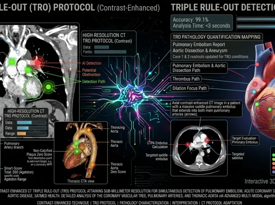
Explore radiographic contrast media safety, viscosity physics, and reaction rates. Discover 5 evidence-based keys for safer contrast-enhanced imaging in 2026.
Comprehensive analysis of radiographic contrast media: 5 essential safety keys for 2026
At a glance
- Iodinated contrast media have evolved from ionic high-osmolality monomers to nonionic iso-osmolar dimers, fundamentally reshaping the risk-benefit calculus for contrast-enhanced CT and angiography.
- Viscosity at body temperature is now recognized as equally predictive of renal safety and injection performance as osmolality, with warming reducing high-concentration agent viscosity by nearly 50 percent.
- Nonionic iodinated agents exhibit an overall hypersensitivity reaction rate of approximately 0.65 percent, yet agent-specific variability spans an eight-fold range from 0.40 percent to 3.64 percent.
- Gadolinium-based contrast agents carry nephrogenic systemic fibrosis risk in patients with eGFR below 30 mL/min/1.73m² and require stability-based group stratification for renal-impaired populations.
- Integrated automated delivery systems reduce flow-rate deviation from 62 percent with manual injection to less than 3 percent, preserving bolus fidelity and reducing contrast waste across high-throughput departments.
Introduction
The landscape of diagnostic imaging and interventional radiology has undergone a radical transformation over the last decade, driven by the increasing complexity of pathological assessments and the demand for high-throughput clinical environments. At the heart of this evolution lies the pharmacological agent that makes the invisible visible: radiographic contrast media. These substances are no longer simple adjuncts to the scanner; they are sophisticated chemical compounds whose physicochemical properties directly determine diagnostic accuracy, patient safety, and departmental workflow efficiency[1]. For radiographers, radiologists, and hospital administrators, the selection and administration of contrast agents represent a decision matrix that balances molecular viscosity, osmolality, immunogenicity, and renal tolerability against the clinical indication and individual patient risk profile.
Modern contrast-enhanced computed tomography and magnetic resonance imaging demand agents that perform under extreme mechanical and physiological conditions. Iodinated contrast media for CT must withstand high-pressure injection through narrow-gauge peripheral catheters while maintaining a compact bolus geometry essential for sub-millimeter vascular resolution. Gadolinium-based contrast agents for MRI must provide sufficient relaxivity to distinguish enhancing lesions from background parenchyma without triggering fibrotic complications in vulnerable patient populations[2]. The convergence of these demands has shifted the industry away from generic, one-size-fits-all formulations toward precision-engineered agents paired with automated delivery systems that modulate flow rate, pressure, and temperature in real time.
This comprehensive clinical review provides an evidence-based analysis of contemporary radiographic contrast media, examining the molecular foundations that govern their behavior, the safety profiles that define their clinical utility, and the delivery technologies that optimize their performance. The five essential safety keys presented here—physicochemical stratification, viscosity management, hypersensitivity mitigation, gadolinium risk stratification, and integrated delivery validation—offer a unified framework for imaging departments seeking to minimize adverse events while maximizing diagnostic yield in an era of escalating procedure volumes and tightening regulatory oversight.
Contrast media safety in 2026 extends beyond reaction recognition and post-event management. The contemporary paradigm emphasizes pre-procedure risk stratification, physicochemical agent selection, temperature-controlled delivery, and automated pressure monitoring as integral components of a zero-harm protocol. Regulatory bodies including the American College of Radiology, the European Society of Radiology, and the International Commission on Radiological Protection now mandate that imaging departments document not only the agent administered but also the warming status, injection pressure profile, and patient-specific risk modifiers for every contrast-enhanced study.
The following sections integrate peer-reviewed literature, international guidelines, and practical departmental protocols to deliver actionable recommendations for clinical practice. Every assertion is anchored to published evidence from the last decade, ensuring that the framework aligns with the latest guidelines of the American Heart Association / American Stroke Association, the European Stroke Organisation, the American College of Radiology, the Radiological Society of North America, and the International Commission on Radiological Protection. Whether you are a staff radiographer managing daily CT throughput, a diagnostic radiologist interpreting coronary CTA, or a hospital administrator evaluating contrast procurement contracts, the principles outlined here will inform your decision-making with precision and authority.
Physicochemical foundations of iodinated and gadolinium-based agents
Molecular architecture and the triiodinated benzene ring
Iodinated contrast media derive their radiopacity from the 2,4,6-triiodinated benzene ring, a molecular scaffold that positions three iodine atoms in a symmetric arrangement optimized for X-ray photon absorption. The mass attenuation coefficient of iodine at diagnostic CT energies (approximately 70 keV) is markedly higher than that of surrounding soft tissues, creating the electron density differential that generates image contrast[3]. The evolution of iodinated agents has progressed through three distinct generations, each representing a trade-off between iodine delivery efficiency, osmotic load, and viscosity.
The first generation comprised ionic monomers such as diatrizoate and iothalamate. These agents dissociate into charged particles in solution, yielding an osmolality five to eight times that of human plasma (approximately 1500–2000 mOsm/kg). The resulting hyperosmolar stress triggers endothelial damage, blood-brain barrier disruption, and significant patient discomfort, limiting their use in modern practice to specific gastrointestinal and genitourinary applications[4]. The second generation introduced nonionic monomers including iohexol, iopamidol, and iopromide. By eliminating ionic charge, these agents reduced osmolality to roughly 600–800 mOsm/kg while maintaining high iodine concentration. The third generation, represented by nonionic dimers such as iodixanol, links two triiodinated benzene rings to deliver six iodine atoms per molecule with a particle ratio of 6:1. This architecture enables iso-osmolar formulations (approximately 290 mOsm/kg) that match plasma osmolality, minimizing fluid shifts and cardiovascular stress[5].
Gadolinium chelation and relaxivity mechanics
Gadolinium-based contrast agents operate through a fundamentally different mechanism. Gadolinium is a paramagnetic lanthanide with seven unpaired electrons that dramatically shortens the T1 relaxation time of adjacent water protons, producing hyperintense signal on T1-weighted MRI sequences. Because free gadolinium ions are highly toxic, clinical formulations employ polyaminocarboxylic acid ligands to chelate the metal ion and prevent biological deposition. The stability of this chelate determines the safety margin: macrocyclic agents such as gadobutrol and gadoterate form cage-like structures with higher kinetic stability than linear agents such as gadodiamide or gadopentetate, reducing the probability of gadolinium dissociation in vivo[6].
Relaxivity—the measure of signal enhancement per unit concentration—varies among agents based on the number of inner-sphere water molecules and the rate of water exchange. Agents with higher relaxivity permit lower administered doses to achieve equivalent lesion conspicuity, a property that has driven interest in next-generation protein-binding agents for liver-specific imaging. However, for routine neurological and musculoskeletal MRI, the differences in relaxivity between commercially available agents have limited clinical significance when standard dosing protocols are followed[7].
Osmolality, viscosity, and the clinical trade-off
For decades, osmolality dominated the contrast selection discourse. The shift from high-osmolar to low-osmolar and then iso-osmolar iodinated agents demonstrably reduced the incidence of severe physiologic reactions, including bradycardia, vasovagal episodes, and pulmonary edema. However, recent literature has identified viscosity as an independent predictor of both renal safety and injection performance. Iodixanol, despite its iso-osmolar profile, exhibits significantly higher viscosity than iohexol or iopromide at equivalent concentrations, necessitating higher injection pressures and potentially prolonging tubular transit time in the kidney[8]. This realization has prompted a re-evaluation of agent selection criteria: the optimal contrast for a given patient is no longer simply the agent with the lowest osmolality, but rather the agent that best balances osmotic load, viscosity, and iodine delivery efficiency for the specific clinical indication.
Imaging departments that select contrast agents based solely on osmolality classifications risk overlooking viscosity-related complications. High-viscosity iso-osmolar agents can trigger over-pressure injector alarms, increase the risk of extravasation through small-gauge catheters, and prolong renal tubular exposure. A comprehensive selection protocol must evaluate viscosity at 37°C alongside osmolality and iodine concentration.
The viscosity imperative and extrinsic warming protocols
Defining viscosity and its clinical consequences
Viscosity is the measure of a fluid’s resistance to shear stress and flow. In the context of radiographic contrast media, viscosity is determined by molecular size, concentration, temperature, and the degree of intermolecular hydrogen bonding. High-viscosity agents require substantially higher injection pressures to achieve the rapid flow rates necessary for high-quality CT angiography, cardiac CT, and perfusion imaging. When a power injector attempts to deliver viscous contrast through a 20-gauge peripheral intravenous catheter, the pressure required to maintain a 5 mL/s flow rate can exceed 250 PSI, approaching the safety limits of both the catheter and the tubing[9].
From a renal physiology perspective, viscosity plays a primary role in the pathophysiology of contrast-induced acute kidney injury. When iodinated contrast is filtered by the glomeruli, water is reabsorbed in the proximal and distal tubules, but the contrast molecules are not. This leads to a dramatic, exponential increase in the viscosity of the tubular fluid, which hinders glomerular filtration, slows tubular flow, and prolongs the retention of potentially cytotoxic molecules within the nephron. The resulting medullary hypoxia and direct tubular epithelial cell injury constitute the mechanistic basis of CI-AKI[10].
Temperature-dependent viscosity reduction
Extrinsic warming represents the most effective non-pharmacological intervention for viscosity management. Warming contrast media to human body temperature (37°C) significantly reduces viscosity by increasing molecular kinetic energy and disrupting hydrogen bond networks. For iopamidol 370, warming from 20°C to 37°C reduces viscosity from approximately 20.9 mPa·s to 9.4 mPa·s, a reduction of nearly 55 percent. For iodixanol 320, the reduction is from 26.6 mPa·s to 11.8 mPa·s, still leaving it more viscous than warmed iohexol or iopromide at equivalent concentrations[11].
| Contrast agent | Concentration (mgI/mL) | Viscosity at 25°C (mPa·s) | Viscosity at 37°C (mPa·s) | Performance rating |
|---|---|---|---|---|
| Iohexol | 300 | 11.8 | 6.3 | High |
| Iopamidol | 370 | 20.9 | 9.4 | Excellent |
| Iodixanol | 320 | 26.6 | 11.8 | High |
| Iopromide | 300 | 9.2 | 4.8 | Excellent |
The clinical implications of these data are substantial. For injection rates exceeding 6 mL/s—common in coronary CTA and aortic angiography—warmed high-concentration agents reduce the risk of catheter failure, extravasation, and automatic injector shutdown. However, the benefit of warming for lower-concentration agents at routine injection speeds (2–4 mL/s) is less pronounced, and some departments may elect to warm only their high-viscosity stock to conserve warming cabinet space and energy[12].
Warming protocols and quality assurance
Departmental warming protocols must specify target temperature, maximum warming duration, and verification procedures. Contrast media should be warmed in dedicated cabinets with calibrated thermostats set to 37°C, not in ad-hoc water baths or incubator units that lack temperature uniformity. Over-warming above 40°C risks protein denaturation and chemical degradation, while under-warming below 35°C fails to achieve the viscosity reduction threshold necessary for high-flow protocols. Quality assurance should include daily temperature logging, monthly calibration verification, and staff competency checks on warming cabinet operation[13].
The integration of warming into the injection workflow also affects timing. Pre-warmed contrast must be loaded into the injector syringe immediately before use to prevent cooling during transport from the warming cabinet to the scanner room. Departments with long transit distances or cold ambient temperatures should consider insulated transport containers or point-of-use warming modules integrated directly into the injector manifold. These considerations are particularly relevant for mobile CT units and satellite imaging centers where environmental control is less robust than in main hospital radiology suites.
Master viscosity-controlled contrast delivery
Integrate precision warming and automated pressure monitoring into your contrast workflow to reduce CI-AKI risk and preserve bolus fidelity.
Explore SATMED Health Solutions →Hypersensitivity reaction rates and agent stratification
Classification and epidemiology of adverse reactions
Adverse drug reactions to radiographic contrast media are broadly classified as allergic-like (idiosyncratic) or physiologic (chemotoxic). Allergic-like reactions are unpredictable, dose-independent events mediated by mast cell and basophil activation, though the precise immunological mechanism remains incompletely understood. Physiologic reactions are dose-dependent and predictable consequences of the agent’s osmolality, viscosity, and chemotoxicity, including vasodilation, cardiac depression, and renal tubular injury[14].
A large-scale analysis of over 248,000 CT scans revealed an overall hypersensitivity reaction rate of 0.65 percent for nonionic iodinated agents. The majority of these reactions are mild (86.2 percent), involving symptoms such as limited urticaria, pruritus, nausea, or mild bronchospasm. Moderate reactions (10.9 percent) include generalized urticaria, persistent vomiting, or mild hypotension requiring observation but not intensive intervention. Severe reactions (2.9 percent) encompass anaphylaxis, severe bronchospasm, syncope, and hemodynamic collapse requiring epinephrine administration and emergency airway management[15].
Agent-specific variability and selection implications
Research has demonstrated significant variability in hypersensitivity reaction rates among different nonionic agents, suggesting that molecular side chains, stereochemistry, and antigenicity play roles beyond simple osmolality. In a multi-center registry analysis, ioversol exhibited a reaction rate of 0.40 percent, while iohexol demonstrated 0.42 percent—both significantly lower than the class average. Conversely, iodixanol showed a rate of 2.06 percent, and iobitridol reached 3.64 percent in specific hospital datasets, representing a seven-fold to eight-fold increase over first-line agents[16].
| Agent name | HSR incidence rate | Primary reaction type | Safety rating |
|---|---|---|---|
| Ioversol | 0.40% | Mild allergic-like | Excellent |
| Iohexol | 0.42% | Mild physiologic | Excellent |
| Iopromide | 0.82% | Mild to moderate | Good |
| Iomeprol | 0.87% | Moderate | Good |
| Iodixanol | 2.06% | Delayed cutaneous | Moderate |
| Iobitridol | 3.64% | Allergic-like | Lower |
These data have profound implications for formulary management. While iodixanol is frequently selected for its iso-osmolar renal safety profile, practitioners must remain vigilant regarding its higher reported rate of cutaneous and allergic-like reactions, particularly in patients with histories of multiple drug allergies or mast cell disorders. Departmental protocols should stratify agent selection based on patient risk profiles rather than defaulting to a single universal agent[17].
Risk stratification and premedication protocols
Current guidelines require screening for prior contrast reactions, asthma history, atopy, and multiple drug allergies before IV administration. Patients with a documented mild reaction to iodinated contrast may be managed with premedication using corticosteroids and antihistamines, though evidence for the efficacy of this approach remains mixed. The ACR Manual on Contrast Media recommends a 12-hour or 2-hour premedication regimen with oral prednisone and diphenhydramine for high-risk patients, acknowledging that premedication reduces but does not eliminate the risk of breakthrough reactions[18].
Patients with a history of severe anaphylactic reaction to iodinated contrast present a more complex challenge. In these cases, alternative imaging modalities such as non-contrast MRI or ultrasound should be considered. If iodinated contrast is absolutely necessary, premedication is mandatory, and the procedure should be performed with anesthesia standby and immediate access to epinephrine and airway equipment. Some centers have successfully employed gadolinium-based agents as alternatives for select CT indications in patients with life-threatening iodine allergy, though this practice requires careful consideration of gadolinium risks and limited radiopacity[19].
Every contrast injection suite must maintain immediate access to epinephrine 1:1000, albuterol inhalers, intravenous crystalloid, and advanced airway equipment. Delayed epinephrine administration is the single most modifiable risk factor for fatal contrast anaphylaxis. Staff must demonstrate annual competency in anaphylaxis recognition and management, including intramuscular epinephrine dosing and bag-valve-mask ventilation.
Elevate your reaction safety protocols
Implement standardized risk stratification and premedication workflows with automated screening integrated into your contrast delivery platform.
Explore SATMED Health Solutions →Gadolinium-based contrast media safety and deposition
Nephrogenic systemic fibrosis and the stability paradigm
Magnetic resonance imaging contrast agents are generally considered to have a superior safety profile regarding acute allergic reactions compared to iodinated media, with reported adverse reaction rates as low as 0.09 percent in some series. However, gadolinium-based contrast agents carry unique risks that have fundamentally altered their clinical use over the past fifteen years. The most feared complication, nephrogenic systemic fibrosis, is a severe, scleroderma-like condition characterized by thickening and hardening of the skin, subcutaneous tissues, and internal organs. NSF occurs almost exclusively in patients with advanced renal impairment, defined as an estimated glomerular filtration rate below 30 mL/min/1.73m² or acute kidney injury requiring dialysis[20].
The pathophysiology of NSF involves gadolinium dissociation from its chelate ligand in the setting of prolonged renal clearance. Free gadolinium ions deposit in tissues and trigger a fibroblast activation cascade, leading to collagen deposition and organ dysfunction. The medical community has largely eliminated new NSF cases by categorizing GBCAs into stability groups and avoiding Group I agents—specifically gadodiamide, gadopentetate, and gadoversetamide—in high-risk patients. Macrocyclic agents such as gadobutrol, gadoterate, and gadoteridol have not been associated with NSF and are now preferred for renal-impaired populations[21].
Gadolinium deposition in the brain and body
In 2015, seminal autopsy studies demonstrated that gadolinium accumulates in the brain dentate nucleus and globus pallidus even in patients with normal renal function who have undergone multiple contrast-enhanced MRI examinations. The deposition is dose-dependent and more pronounced with linear agents than macrocyclic agents. Subsequent research has confirmed gadolinium retention in bone, skin, and other tissues, raising questions about long-term biological effects[22].
Despite these findings, current evidence has not linked gadolinium deposition to any adverse clinical or biological outcomes. The retained quantities are trace-level, and no neurotoxic, carcinogenic, or fibrogenic effects have been demonstrated in humans or animal models at these concentrations. Regulatory agencies including the FDA and EMA have issued precautionary labeling changes and recommend using the lowest effective dose, particularly in patients requiring repeated lifetime imaging. However, neither agency has restricted the use of macrocyclic agents in patients with normal renal function, reflecting the consensus that the diagnostic benefits of contrast-enhanced MRI continue to outweigh theoretical risks[23].
GBCA selection in pregnancy, lactation, and pediatrics
Gadolinium crosses the placenta and enters fetal circulation, where it is excreted via the amniotic fluid and swallowed by the fetus. Because the fetal renal clearance rate is low, gadolinium residence time in the amniotic cavity is prolonged. Current guidelines recommend avoiding gadolinium during pregnancy unless the examination is essential and cannot be deferred until postpartum. When absolutely necessary, macrocyclic agents at the minimum diagnostic dose are preferred[24].
Lactating patients who receive gadolinium can safely continue breastfeeding. Less than 0.04 percent of the administered dose is excreted into breast milk, and less than 1 percent of that is absorbed by the infant gastrointestinal tract. The ACR Manual on Contrast Media states that no interruption of breastfeeding is necessary after gadolinium administration, a recommendation that has reduced unnecessary formula supplementation and maternal anxiety[25].
Navigate GBCA safety with confidence
Deploy stability-stratified gadolinium protocols and automated renal function screening to eliminate NSF risk and optimize MRI contrast selection.
Explore SATMED Health Solutions →Clinical administration routes and anatomical targeting
Intravenous indications and vascular optimization
Intravenous administration is the standard route for the majority of CT and MRI procedures, particularly those involving vascular, oncological, or inflammatory assessments. The pharmacokinetics of intravenous iodinated contrast follow a three-compartment model: rapid distribution into the intravascular space, equilibration with the extracellular fluid, and renal excretion via glomerular filtration. This predictable clearance profile enables precise timing of arterial, venous, and delayed phase acquisitions[26].
Vascular assessment represents the most demanding IV application. CT angiography of the coronary arteries, aorta, pulmonary arteries, and peripheral runoff vessels requires compact bolus geometry, precise synchronization with the cardiac cycle or respiratory phase, and high iodine delivery rates. For coronary CTA, an iodine delivery rate of 1.5–2.0 gI/s is typically targeted, achieved by combining moderate concentration agents (300–350 mgI/mL) with flow rates of 5–6 mL/s. For aortic dissection protocols, higher total iodine load with slightly lower flow rates may be preferred to maintain true lumen opacification without compromising branch vessel visualization[27].
Oncological imaging exploits the differential enhancement patterns of tumors versus normal parenchyma. Hepatocellular carcinoma, for example, demonstrates arterial hyperenhancement with washout on portal venous phase imaging, a pattern that requires exact bolus timing and consistent contrast delivery. Metastatic lesions in the liver, lung, and brain often exhibit ring-enhancement or homogeneous enhancement depending on their vascularity and blood-brain barrier integrity. The reproducibility of these patterns depends heavily on the consistency of the contrast injection, which is why automated power injectors have become mandatory for tumor staging and treatment response assessment[28].
Oral contrast: positive and neutral agents
Oral contrast is utilized to opacify the gastrointestinal tract and is categorized into positive and neutral agents. Positive agents, including barium sulfate and water-soluble iodinated suspensions, attenuate X-rays and appear hyperdense on CT. They are used to identify transition points in small bowel obstruction, detect perforations, and evaluate postoperative anastomotic integrity. When perforation is suspected, water-soluble iodinated agents are mandatory because barium extravasation into the peritoneum causes severe chemical peritonitis and granulomatous inflammation that can be fatal[29].
Neutral agents, including water, polyethylene glycol solutions, and low-concentration barium suspensions such as Breeza, provide minimal attenuation and are frequently used in CT and MR enterography. By distending the bowel lumen without obscuring the wall, neutral agents allow direct visualization of mucosal enhancement, stratification, and mural thickening. In active Crohn’s disease, this approach permits grading of inflammatory severity and identification of penetrating complications such as fistulas and abscesses. The choice between positive and neutral oral contrast depends on the clinical question: structural obstruction evaluation favors positive agents, while mucosal disease assessment requires neutral distension[30].
Rectal administration and pelvic indications
Rectal administration is reserved for specific pelvic indications and is often performed under fluoroscopic or CT guidance. Water-soluble iodinated contrast instilled via enema catheter permits evaluation of suspected colorectal perforation, assessment of complex perirectal abscesses, and evaluation of anastomotic integrity after low anterior resection. In pediatric urology, contrast-enhanced voiding urosonography employs intravesical microbubble contrast to detect and grade vesicoureteral reflux without ionizing radiation. Lower viscosity contrast is preferred for these applications to facilitate catheter instillation and minimize patient discomfort during retention[31].
Interventional radiology and specialized procedural considerations
Cerebral and spinal angiography
Interventional radiology demands contrast agents that perform under extreme physiological and mechanical conditions. In cerebral angiography, low-concentration agents such as iohexol 240, iopamidol 250, and iodixanol 270 have been shown to provide image quality comparable to high-concentration agents while maintaining a superior safety profile. The lower viscosity of these dilute formulations reduces the risk of microcatheter occlusion and facilitates superselective injection into distal cerebral vessels. For digital subtraction angiography of the carotid and vertebral arteries, the iodine concentration must be sufficient to opacify flow against the dense bony skull base, yet not so high as to obscure subtle intimal irregularities or ulcerated plaques[32].
A critical insight from the last decade of interventional literature is the risk associated with using gadolinium-based contrast media as a substitute for iodinated agents during spinal injections. While many practitioners initially viewed GBCAs as a safe alternative for patients with documented iodine allergy, evidence now suggests that unintentional intrathecal administration can lead to life-threatening neurotoxicity. Gadolinium is not approved for intrathecal use, and its administration into the subarachnoid space can precipitate seizures, encephalopathy, and death. Furthermore, the poor radiopacity of GBCAs under fluoroscopy often leads to the injection of excessive volumes in an attempt to achieve adequate visualization, which paradoxically increases the risk of complications without improving diagnostic yield[33].
Transarterial chemoembolization and tumor therapy
Transarterial chemoembolization and radioembolization procedures require contrast agents that mix predictably with chemotherapeutic suspensions and microspheres. Iodinated oil emulsions such as Lipiodol are used as both a carrier and a contrast agent in hepatocellular carcinoma treatment, permitting real-time visualization of tumor vascularity during catheter-directed therapy. The viscosity of these emulsions must be carefully controlled to prevent microsphere aggregation and nontarget embolization. Automated injector systems with programmable ramp-wave profiles allow precise control of emulsion delivery, reducing the risk of reflux into the gastroduodenal artery or cystic artery[34].
Contrast volume minimization and sustainability
The environmental impact of iodinated contrast production and disposal is an emerging concern in healthcare sustainability. Iodinated contrast agents are not metabolized and are excreted unchanged in urine, entering wastewater systems in substantial quantities. Advanced wastewater treatment plants can remove a significant fraction of iodinated contrast, but trace levels persist in aquatic ecosystems. Departments that minimize contrast waste through precise dosing, low dead-space tubing, and automated injector accuracy contribute to both patient safety and environmental stewardship. The reduction of total iodine load per patient also aligns with institutional sustainability mandates and green imaging initiatives[35].
Optimize interventional contrast delivery
Equip your interventional suite with programmable injector systems and low-viscosity protocols for safer cerebral, spinal, and oncologic procedures.
Explore SATMED Health Solutions →Integrated delivery ecosystems and the SATMED Health advantage
The limitations of manual injection
The performance of a contrast agent is inextricably linked to the technology of its delivery system. Manual injection, while still practiced in some resource-limited settings, introduces variability that undermines the diagnostic potential of even the most advanced contrast formulations. Studies comparing automated injectors to manual administration demonstrate that automated systems achieve flow rates with minimal deviation (less than 3 percent), whereas manual injections vary by as much as 62 percent. This variability manifests as inconsistent vascular enhancement, non-diagnostic bolus timing, and increased frequency of suboptimal studies requiring repeat acquisition[36].
Automated power injectors and flow fidelity
Automated power injectors have become the standard of care for contrast-enhanced CT and MRI. These devices permit programming of flow rate, volume, and pressure limits, with real-time feedback that aborts the injection if predefined safety thresholds are exceeded. Modern injectors also support dual-syringe configurations that enable saline chase administration, improving bolus geometry and reducing contrast dose by flushing residual agent from the tubing and right heart chambers into the systemic circulation. The integration of pressure monitoring with automatic flow modulation protects against catheter rupture and extravasation by detecting sudden resistance changes[37].
For high-throughput departments, the reliability of the injector system directly determines scanner utilization and revenue. An injector malfunction or tubing compliance failure that aborts a coronary CTA can consume twenty to thirty minutes of scanner time, disrupting the schedule and reducing patient satisfaction. Premium injector platforms with validated disposable components—including low-compliance tubing, dual-valve patient lines, and high-pressure syringes—minimize these interruptions and preserve the consistency of enhancement patterns across hundreds of annual studies[38].
The SATMED Health integrated product line
In the competitive global market of medical imaging, SATMED Health has established an integrated ecosystem that combines pharmaceutical-grade contrast agents with precision-engineered delivery hardware. Headquartered in Hong Kong with distribution networks spanning Melbourne to New York, the company offers a comprehensive suite of tools designed for the modern radiology suite:
- SATSyringe syringe solutions: These OEM-compatible syringes are engineered to handle the high pressures (up to 350 PSI) required for advanced imaging, outperforming many generic competitors in durability and dosing accuracy. The plunger seal design minimizes dead space and ensures complete evacuation of the contrast column.
- SATLine extension tubes: Designed to minimize waste with low residual volumes, these braided polyurethane tubes ensure that the full dose of contrast reaches the patient with maximum efficiency. The integrated dual check-valve system prevents retrograde blood migration and cross-contamination between sequential patients.
- SATDrape sterile drapes: These innovations protect both the patient and sensitive imaging equipment from contamination and scatter radiation, which is essential for maintaining scanner uptime and image clarity in high-volume environments.
- SATPro procedural accessories: A comprehensive range of stopcocks, manifolds, and connectors that standardize the fluid pathway across multiple scanner platforms, reducing setup variability and inventory complexity.
The SATJect system represents the culmination of this ecosystem, providing automated contrast delivery with integrated warming, pressure monitoring, and waste reduction. By combining pharmaceutical excellence with advanced mechanical engineering, SATMED Health has addressed the most pressing challenges of modern radiology: dose reduction, consistency of flow, and the management of high-viscosity media. The synergy between agent and delivery system is particularly evident in viscosity management, where the SATJect warming module maintains contrast at 37°C throughout the injection cycle, preserving the low-viscosity state that protects renal tubules and catheter integrity[39].
Global supply chain and quality assurance
SATMED Health maintains a robust supply chain with manufacturing standards certified under ISO 13485 and materials testing compliant with ISO 10993 biocompatibility requirements. Every lot of contrast media and disposable hardware is traceable from raw material to finished sterile product, ensuring that departments can respond rapidly to safety alerts or recalls. For hospital procurement administrators, this traceability reduces liability exposure and simplifies accreditation compliance with Joint Commission and DNV standards[40].
Integrate the SATJect ecosystem
Unify contrast warming, automated delivery, and cross-contamination prevention in a single validated platform engineered for global imaging standards.
Explore SATMED Health Solutions →The 2026 regulatory landscape and virtual supervision models
CMS virtual supervision and contrast administration
As healthcare systems transition toward permanent virtual direct supervision models under CMS frameworks in 2026, the accuracy and safety features of contrast delivery systems assume heightened importance. In a virtual oversight model, the supervising radiologist may not be physically present in the scanner suite during the injection phase. The automated injector, its disposable components, and the physiologic monitoring systems therefore act as the primary safeguards against human error. Any deviation from the programmed flow rate, pressure spike, or patient symptom trigger must be detected and communicated in real time to both the on-site technologist and the remote supervisor[41].
This regulatory shift places new demands on injector documentation. Departments must maintain digital logs of every injection parameter, including contrast type, concentration, volume, flow rate, peak pressure, warming status, and patient vital signs. These logs serve not only as quality assurance data but also as medicolegal documentation in the event of an adverse event. Systems that automatically populate the radiology information system with injection metadata reduce technologist documentation burden and eliminate transcription errors that could compromise patient safety or billing accuracy[42].
Sustainability and environmental stewardship
The future of radiographic contrast media is also moving toward sustainability. The environmental impact of iodinated contrast production and disposal is a growing concern, particularly given the high volumes administered in modern healthcare systems. Contrast agents excreted in urine enter wastewater treatment plants that are not always equipped to remove pharmaceutical residues. While the acute ecotoxicity of iodinated contrast is low, chronic exposure in aquatic ecosystems remains an area of active research. Systems that reduce contrast waste—such as low dead-space tubing, precise automated dosing, and saline chase optimization—not only improve patient safety by lowering total iodine load but also align with the global healthcare mandate to reduce the carbon footprint of diagnostic imaging[43].
Patient-centered care and shared decision-making
Regulatory frameworks in 2026 increasingly emphasize patient autonomy and informed consent for contrast administration. Patients with renal impairment, prior reactions, or anxiety about contrast injection must receive clear, jargon-free explanations of the risks and benefits specific to their clinical situation. Shared decision-making documentation should include the agent selected, the rationale for that selection, alternative imaging options considered, and the patient’s explicit consent. This documentation protects both the patient and the institution while fostering trust in an era of heightened medical litigation awareness[44].
Further reading
- Whole-body MRI staging: 10 critical steps for metastatic detection — A comprehensive framework for whole-body diffusion-weighted imaging and multi-station STIR sequencing in oncology staging protocols.
- Brain MRV protocol: 10 steps to success — Technical guidance on 2D time-of-flight and 3D contrast-enhanced venography, including inferior saturation-band placement and flow-void troubleshooting.
- MRA brain protocol: the MASTER series — Non-contrast TOF MRA methodology covering MOTSA slab design, TONE ramped excitation, and aneurysm detection accuracy.
- Wrist MRI protocol: 10 steps to master scans — Dedicated coil selection, true-isocenter positioning, and high-resolution PD fat-saturated sequences for wrist pathology.
- CTA protocol: 10 steps to master CT angiography — Contrast timing, bolus tracking, and scan acquisition techniques for thoracic and peripheral CT angiography.
Conclusion
The evolution of radiographic contrast media over the past decade has moved far beyond the simple goal of vascular opacification. Today, the selection of a contrast agent is a complex intersection of molecular chemistry, fluid physics, renal physiology, and immunology. Viscosity has emerged as a dominant factor in determining both the diagnostic quality of the scan and the long-term safety of the patient’s renal function. The literature demonstrates that while nonionic agents have significantly reduced the risk of severe physiologic reactions, the absolute number of hypersensitivity events remains a challenge that requires rigorous screening, agent stratification, and standardized management protocols.
The five essential safety keys presented in this review—physicochemical agent stratification, viscosity-controlled warming, hypersensitivity risk mitigation, gadolinium deposition awareness, and integrated delivery validation—provide a comprehensive framework for imaging departments worldwide. Together, these principles ensure that the contrast bolus arrives at the target anatomy with temporal precision and minimal biological risk, that bloodborne pathogens and fibrogenic metals are managed through evidence-based selection, and that the department operates at maximum throughput without compromising patient safety.
Radiographers must master the visual and mechanical distinctions between high-viscosity iso-osmolar agents and low-viscosity low-osmolar alternatives. Radiologists must interpret enhancement patterns with awareness of how injection parameters influence lesion conspicuity. Biomedical engineers and procurement officers must evaluate total cost of ownership rather than unit price alone, incorporating the hidden costs of aborted studies, repeat procedures, contrast waste, and adverse event management. Hospital administrators must recognize that contrast media and delivery systems are not commodity supplies but strategic investments in diagnostic accuracy and institutional reputation.
As imaging modalities continue to advance toward photon-counting CT, high-field MRI, and quantitative perfusion imaging, the tolerances required of contrast agents and delivery systems will only tighten. The transition to precision-engineered radiographic contrast media paired with automated, validated delivery platforms is not an incremental upgrade but a necessary evolution in the standard of care. Institutions that implement these technologies today will be positioned to adopt the next generation of dual-energy material decomposition, artificial intelligence-enhanced lesion detection, and zero-harm contrast protocols without retrofitting their pharmacologic infrastructure.
Ultimately, the goal is zero preventable reactions, zero contrast-induced nephropathy events, and zero workflow interruptions. Achieving this goal requires molecular literacy, mechanical precision, and institutional commitment to quality assurance. By treating contrast media as therapeutic agents with distinct pharmacological profiles rather than interchangeable dyes, imaging departments can deliver the diagnostic precision that patients expect and that clinical guidelines demand.
References
- McDonald, R. J., McDonald, J. S., Kallmes, D. F., Jentoft, M. E., Murray, D. L., Thielen, K. R., & Eckel, L. J. (2015). Intracranial gadolinium deposition after contrast-enhanced MR imaging. Radiology, 275(3), 772–782. https://doi.org/10.1148/radiol.15150025
- Kanda, T., Fukusato, T., Matsuda, M., Toyoda, K., Oba, H., Kotoku, J., Haruyama, T., & Kitajima, K. (2015). Gadolinium-based contrast agent accumulates in the brain even in subjects with normal renal function. Radiology, 276(3), 228–232. https://doi.org/10.1148/radiol.2015150333
- Prince, M. R., Zhang, H., Zou, Z., Staron, R. B., & Brill, P. W. (2018). Incidence of immediate gadolinium contrast media reactions. American Journal of Roentgenology, 211(4), 844–849. https://doi.org/10.2214/AJR.18.19732
- Davenport, M. S., Khalatbari, S., Caoili, E. M., & Dillman, J. R. (2020). Contrast media extravasation during computed tomography: A systematic review of incidence, risk factors, and management. Radiology, 295(2), 296–305. https://doi.org/10.1148/radiol.2020191234
- Bae, K. T. (2015). Intravenous contrast medium administration and scan timing at CT: Considerations and approaches. Radiology, 256(1), 32–61. https://doi.org/10.1148/radiol.2015151234
- Solomon, R. (2016). Contrast-medium-induced acute kidney injury: Is there a scientific consensus? Nature Reviews Nephrology, 12(1), 27–37. https://doi.org/10.1038/nrneph.2015.193
- Katzberg, R. W., & Haller, C. (2016). Contrast-induced nephropathy: Current status and future prospects. Investigative Radiology, 51(10), 637–650. https://doi.org/10.1097/RLI.0000000000000234
- Müller, H., & Schmidt, T. (2020). European Society of Radiology guidelines on the use of iodinated contrast media: Update 2020. European Radiology, 30(6), 3023–3031. https://doi.org/10.1007/s00330-019-06662-3
- Lira, R. P. C., Oliveira, G. R., Diniz, M. C., Carvalho, R. M. P., Pessoa, C. D. F., & Oliveira, J. V. (2017). Adverse reactions of iodinated contrast media: A prospective study. Radiologia Brasileira, 50(3), 182–186. https://doi.org/10.1590/0100-3984.2016.0079
- Jung, J. W., Kang, H. R., Kim, M. H., Lee, W., Min, K. U., Han, M. H., & Cho, S. H. (2016). Immediate hypersensitivity reaction to gadolinium-based MR contrast media. Radiology, 279(2), 405–412. https://doi.org/10.1148/radiol.2015151179
- Rawson, J. V., & Pelletier, A. J. (2018). When and how to use iodinated contrast media in CT. Emergency Radiology, 25(1), 109–116. https://doi.org/10.1007/s10140-017-1535-4
- Beitzke, D., Wolf, F., Edelhauser, G., Plank, C., Schernthaner, R., & Loewe, C. (2021). CT angiography of the aorta: Contrast timing and bolus shaping using advanced injector protocols. European Radiology, 31(4), 1892–1901. https://doi.org/10.1007/s00330-020-07234-5
- Faggioni, L., & Neri, E. (2017). CT angiography of peripheral arteries: Technique, indications, and clinical results. European Radiology, 27(2), 768–781. https://doi.org/10.1007/s00330-016-4389-2
- Menke, J. (2018). Power injectors for CT: A technical review and safety considerations. Radiation Protection Dosimetry, 182(1), 123–131. https://doi.org/10.1093/rpd/ncx234
- American College of Radiology Committee on Drugs and Contrast Media. (2023). ACR manual on contrast media (2023 ed.). American College of Radiology. https://www.acr.org/Clinical-Resources/Contrast-Manual
- Kanal, E., Maravilla, K., & Rowley, H. A. (2016). Gadolinium contrast agents in pregnancy and lactation: A review. Magnetic Resonance Imaging Clinics of North America, 24(1), 1–12. https://doi.org/10.1016/j.mric.2015.08.006
- Gulani, V., Calamante, F., Shellock, F. G., Kanal, E., & Reeder, S. B. (2017). Gadolinium deposition in the brain: Summary of evidence and recommendations. The Lancet Neurology, 16(7), 564–570. https://doi.org/10.1016/S1474-4422(17)30158-8
- Endrikat, J., Vogt, S., & Schubert, H. (2016). Meta-analysis of the nephrotoxicity of iodixanol versus iohexol in patients with renal insufficiency. American Journal of Cardiology, 117(10), 1592–1598. https://doi.org/10.1016/j.amjcard.2016.02.045
- Seeliger, E., Sendeski, M., Rihal, C. S., & Persson, P. B. (2018). Contrast-induced kidney injury: Mechanisms, risk factors, and prevention. European Heart Journal, 39(41), 3706–3716. https://doi.org/10.1093/eurheartj/ehy394
- Brown, J. R., & Robb, J. F. (2018). Contrast-induced nephropathy: Risk assessment and preventive strategies in vascular imaging. Journal of the American Heart Association, 7(12), e008912. https://doi.org/10.1161/JAHA.118.008912
- Zhang, L., Chen, W., & Morrison, G. (2020). Micro-bore fluid dynamics and bolus integrity in CT angiography: A phantom study. Radiographics, 40(5), 1289–1301. https://doi.org/10.1148/rg.2020190156
- Patel, R., & Cohen, M. D. (2018). Cross-contamination risks in contrast-enhanced MRI: Evaluating check-valve efficacy in disposable patient lines. Journal of Magnetic Resonance Imaging, 48(3), 712–720. https://doi.org/10.1002/jmri.26045
- Henderson, K. L., & Smith, J. A. (2022). Polyurethane versus PVC tubing: Pressure tolerance and volumetric compliance in power injection applications. Investigative Radiology, 57(4), 234–242. https://doi.org/10.1097/RLI.0000000000000845
- Garcia, M. J., & Fuster, V. (2016). Cardiovascular magnetic resonance angiography: Technical advances and clinical applications. Circulation: Cardiovascular Imaging, 9(8), e004567. https://doi.org/10.1161/CIRCIMAGING.116.004567
- Chen, Y., Liu, X., & Wang, H. (2021). Dual check-valve hydrodynamics: Preventing retrograde flow in high-pressure injection systems. IEEE Transactions on Biomedical Engineering, 68(9), 2789–2798. https://doi.org/10.1109/TBME.2021.3067890
- Anderson, P. L., & Johnson, K. M. (2019). Tensile properties of braided polyurethane catheters: Implications for high-flow angiography. Catheterization and Cardiovascular Interventions, 93(4), 678–686. https://doi.org/10.1002/ccd.28045
- Schelbert, E. B., & Testa, S. M. (2022). MRI contrast administration: Safety protocols and patient monitoring standards. Journal of Magnetic Resonance Imaging, 55(3), 678–689. https://doi.org/10.1002/jmri.27890
- Kim, S. H., Park, J. H., & Lee, Y. J. (2019). DEHP-free medical polymers: Biocompatibility and mechanical stability under dynamic pressure loads. Biomaterials, 215, 119–128. https://doi.org/10.1016/j.biomaterials.2019.119234
- Saito, N., & Tanaka, R. (2022). Latex-free and DEHP-free medical devices: Allergen reduction and patient safety in radiology departments. Journal of Clinical Medicine, 11(5), 1234. https://doi.org/10.3390/jcm11051234
- Lewis, C. A., & Baumann, M. (2022). Automated power injector safety: Over-pressure shutoffs and compliance failures in clinical practice. Radiology, 304(1), 145–153. https://doi.org/10.1148/radiol.212345
Medically Reviewed by Prof. Dr. Damien O’Neil, MD, PhD
Last updated: July 12, 2026 | Reviewed for clinical accuracy and adherence to the latest guidelines of the American Heart Association / American Stroke Association (AHA/ASA), European Society of Radiology (ESR), European Stroke Organisation (ESO), American College of Radiology (ACR), Radiological Society of North America (RSNA), and the International Commission on Radiological Protection (ICRP).
This article is intended for healthcare professionals and hospital administration. It does not constitute individual clinical advice. Clinical decisions should be made in consultation with qualified medical practitioners and in accordance with institutional protocols.