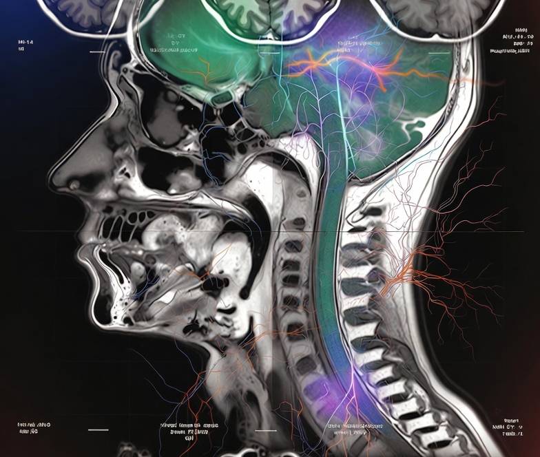
Explaining the mechanical physics behind loud MRI knocking, thumping, and humming sounds, magnetic gradient coils, and mandatory hearing protection safety.
Why Do MRI Scans Make Loud Thumping Sounds?
At a glance
- MRI noise comes from gradient coils vibrating inside the powerful static magnetic field via the Lorentz force.
- Noise levels range from 65 to 110 decibels depending on sequence and field strength—comparable to a chainsaw or rock concert.
- Every imaging facility mandates hearing protection: foam earplugs or MRI-safe noise-reducing headphones.
- Different MRI sequences produce distinct sound patterns because each switches gradient coils at unique frequencies.
- Modern quiet MRI technologies and advanced gradient designs are reducing acoustic noise by up to 80%.
Why are MRI scans so loud? If you have ever had a magnetic resonance imaging scan, you know the experience is accompanied by a symphony of knocking, thumping, buzzing, and humming sounds that can feel startling at first. Many patients describe the noise as resembling a jackhammer, a washing machine with bricks inside, or even a techno music beat. While these sounds are completely normal and expected, understanding why they happen—and how imaging centers protect your hearing—can transform anxiety into informed confidence.
Clinical context: The American College of Radiology (ACR) and the International Electrotechnical Commission (IEC) have established strict acoustic noise limits for MRI scanners. All accredited imaging facilities must provide adequate hearing protection and monitor noise exposure to prevent temporary or permanent hearing damage.
Nearly 52,000 patients search online every month for explanations of MRI noise and hearing protection. This guide breaks down the physics behind the sound, explains why each sequence has its own unique rhythm, and shows you exactly how technologists keep your ears safe during every scan.
🚀 Elevate Your Department's Imaging Standards
Access premium interventional radiology consumables and safety solutions designed for modern cath labs and imaging suites.
Explore SATMED Health Solutions →The physics of gradient coil vibration
The loud knocking noises in an MRI scanner originate from a fundamental principle of electromagnetism called the Lorentz force. To understand this, it helps to know a little about how MRI creates images.
What gradient coils do
An MRI scanner contains three sets of gradient coils—one for each spatial direction (X, Y, and Z). These coils are essentially loops of wire wrapped around the inside of the scanner bore. Their job is to create small, rapidly changing magnetic fields that superimpose on the scanner's main static magnetic field. This allows the computer to determine where each signal is coming from inside your body, building the final image slice by slice.
The Lorentz force in action
When electrical current pulses through a gradient coil, the coil sits inside the scanner's powerful static magnetic field (typically 1.5 or 3.0 Tesla). According to the Lorentz force law, any electrical conductor carrying current inside a magnetic field experiences a physical force. This force causes the gradient coils to vibrate rapidly—expanding and contracting thousands of times per second—against their mechanical mountings. That mechanical vibration is what you hear as the loud knocking and thumping.
Diagnostic pearl: The louder the sequence, the more rapidly the gradient coils are switching. Fast sequences like echo-planar imaging (EPI) used in functional MRI and diffusion-weighted imaging produce the most intense acoustic noise because they require the fastest gradient switching rates.
Eddy currents and secondary vibrations
The primary sound comes from the gradient coils themselves, but secondary vibrations also contribute. Rapid magnetic field changes induce eddy currents in nearby conductive materials—such as the scanner's metal frame, cryostat, and even the patient table. These eddy currents create their own small magnetic fields, which interact with the main field and produce additional vibrations, adding layers of humming and buzzing to the acoustic signature.
Why different sequences sound different
One of the most fascinating aspects of MRI acoustics is that each imaging sequence produces its own distinct sound pattern. If you pay attention, you will notice that the rhythm, pitch, and intensity change multiple times during a single exam. Here is why:
- T1-weighted sequences: Typically produce a slower, rhythmic knocking pattern with moderate intensity. These sequences use relatively simple gradient switching to highlight anatomical structure.
- T2-weighted sequences: Often sound faster and more continuous, with a higher-pitched buzzing quality. The longer echo times require more complex gradient modulation.
- Diffusion-weighted imaging (DWI): Produces some of the loudest, most intense knocking sounds. DWI requires very strong, rapidly switched gradient pulses to measure the random motion of water molecules in tissue.
- Echo-planar imaging (EPI): Creates a rapid-fire machine-gun-like staccato. EPI reads an entire image plane after a single radiofrequency pulse by oscillating gradients at extreme speeds.
- Fluid-attenuated inversion recovery (FLAIR): Begins with a brief silence during the inversion pulse, followed by a patterned knocking sequence as the gradients engage.
Fun fact: Some creative patients and technologists have noted that certain MRI sequences sound remarkably like specific musical genres. EPI sequences have been compared to industrial techno, while spin-echo sequences resemble a slow mechanical drumbeat.
💉 Optimise Your Cath Lab Workflow
SATLine consumables deliver consistent performance across diagnostic and interventional procedures. Reduce waste and standardise inventory.
Discover SATLine Products →How loud is an MRI? Decibel comparisons
Understanding MRI noise levels in context helps patients appreciate why hearing protection is non-negotiable. Sound intensity is measured in decibels (dB) on a logarithmic scale, meaning every 10 dB increase represents a tenfold increase in acoustic power.
Typical MRI noise levels by scanner type
- 1.5T scanners: 65–100 dB depending on sequence
- 3.0T scanners: 85–110 dB depending on sequence
- 7.0T research scanners: Can exceed 120 dB without protection
Real-world decibel comparisons
- Normal conversation: ~60 dB
- Busy traffic: ~70 dB
- Vacuum cleaner: ~80 dB
- Average MRI sequence: ~90 dB
- Loudest MRI sequences (3.0T EPI): ~110 dB
- Rock concert / chainsaw: ~110 dB
- Jet engine at takeoff: ~140 dB
Hearing safety threshold: The Occupational Safety and Health Administration (OSHA) recommends that unprotected exposure to noise above 85 dB be limited to 8 hours per day. At 100 dB, safe exposure drops to just 15 minutes. A single loud MRI sequence can reach these levels in seconds, which is why hearing protection is mandatory.
Hearing protection: what works and what does not
Every accredited MRI facility is required to provide adequate hearing protection for all patients and staff inside the scan room. Here is what you can expect and why it matters:
Foam earplugs
Disposable foam earplugs are the most common form of protection. When inserted correctly—rolled into a thin cylinder, inserted deep into the ear canal, and held in place for 30 seconds while they expand—they can reduce noise exposure by 20–30 dB. This brings even the loudest 110 dB sequences down to a safe 80–90 dB range.
MRI-safe noise-reducing headphones
Many centers offer specialized headphones that fit over foam earplugs. These headphones contain no ferromagnetic metal and are designed specifically for the MRI environment. They serve two purposes: additional noise reduction (another 10–15 dB) and the ability to pipe in music or verbal instructions from the technologist.
What you cannot bring
Standard consumer headphones, AirPods, and wired earbuds are strictly prohibited inside the MRI scan room. They contain metal components that can become dangerous projectiles in the strong magnetic field, and their electronic circuits can interfere with image quality.
Best practice: The most effective protection combines both foam earplugs and MRI-safe headphones. This dual-layer approach can achieve noise reduction of 30–40 dB, making even the loudest sequences feel comparable to normal conversation.
🛡️ Protect Your Procedures with SATDrape
Advanced sterile draping solutions designed for interventional radiology and cath lab environments.
Explore SATDrape Range →Quiet MRI technology: the future
MRI manufacturers have invested heavily in acoustic noise reduction over the past decade. Several engineering innovations are already making scans significantly quieter:
- Gradient coil redesign: Newer scanners use vacuum-encased gradient coils and active vibration damping systems that absorb mechanical energy before it becomes sound.
- Silent scan sequences: Manufacturers like Siemens ("Silent MRI"), GE ("SilentScan"), and Philips ("ComforTone") have developed proprietary pulse sequences that spread gradient switching over longer time periods, reducing peak noise by up to 80% without sacrificing image quality.
- Sound-dampening enclosures: Advanced scanner housing materials and acoustic insulation around the bore absorb and deflect sound waves before they reach the patient.
- Variable density sampling: Some modern sequences use compressed sensing algorithms that require fewer gradient switches overall, naturally reducing acoustic output.
Clinical impact: Quieter scans are not just about comfort. Reduced noise lowers patient anxiety and motion, which improves image quality. For pediatric and claustrophobic patients, silent sequences can mean the difference between a successful scan and a failed appointment.
What patients can do to cope
Beyond hearing protection, here are practical strategies to make the acoustic experience of your MRI more manageable:
- Request music: If your facility offers MRI-safe headphones with music, bring a playlist of calming songs. Familiar music transforms the noise from a source of anxiety into background ambiance.
- Focus on the rhythm: Some patients find it helpful to mentally "conduct" the sounds, treating the sequence changes like movements in a symphony. This cognitive reframing reduces perceived stress.
- Use breathing techniques: Slow, deep breathing activates your parasympathetic nervous system. Try inhaling for 4 counts, holding for 4, and exhaling for 6.
- Know the timeline: Ask your technologist how many sequences are planned and how long each lasts. Most individual sequences run 2–5 minutes. Knowing there is a break coming helps you endure the noise.
- Close your eyes and visualize: Combine an eye mask with mental imagery of a calm, open place. Removing both visual and auditory context helps your brain disengage from the scanner environment.
Patient insight: Many repeat MRI patients report that the noise becomes less startling over time. The first scan is always the most surprising. By the second or third scan, most people find the sounds familiar and far less anxiety-inducing.
🔧 Precision Tools for Every Procedure
SATPro interventional kits provide standardised, ready-to-use instrument sets that streamline prep time and reduce errors.
Browse SATPro Kits →SATCare Clinical Calculators
Instant, evidence-based calculations for contrast dosing, radiation exposure, and extravasation risk assessment.
CT & MRI Contrast
Weight-based contrast dosing with eGFR safety checks.
SATDose Radiation
Effective dose estimation and cumulative tracking.
Extravasation Risk
Pre-injection risk stratification for contrast leaks.
Frequently asked questions
Not when proper hearing protection is used correctly. Foam earplugs or noise-canceling headphones reduce MRI noise levels from 100–110 decibels to completely safe, comfortable ranges well below the hearing damage threshold. All accredited facilities mandate hearing protection for every patient.
Each distinct sound pattern corresponds to a different imaging sequence. T1-weighted, T2-weighted, diffusion-weighted, and other sequences each switch gradient coils at unique frequencies and patterns to capture specific tissue properties. That is why you hear rhythms that change throughout the exam.
MRI scans typically produce noise between 65 and 110 decibels depending on the sequence and magnetic field strength. A 3.0T scanner is generally louder than a 1.5T scanner. For comparison, normal conversation is about 60 dB, a vacuum cleaner is 80 dB, and a chainsaw is about 110 dB.
You must use MRI-safe hearing protection provided by the facility. Standard wired headphones contain metal and are not allowed inside the scanner. The imaging center will supply foam earplugs or specially designed non-metallic noise-reducing headphones that are magnet-safe.
The knocking sound is caused by the Lorentz force. When electrical current passes through gradient coils inside the strong static magnetic field, the coils experience a physical force that makes them vibrate rapidly against their mountings. That mechanical vibration is what produces the characteristic knocking and thumping you hear.
No. Different manufacturers (Siemens, GE, Philips, Canon) use different gradient coil designs, which produce slightly different acoustic signatures. Additionally, 3.0T scanners are generally louder than 1.5T scanners, and newer models with quiet-sequence technology can be dramatically quieter than older machines.
Without hearing protection, repeated exposure to MRI noise above 100 dB could theoretically cause hearing damage. However, this risk is eliminated entirely when proper foam earplugs or MRI-safe headphones are used. No cases of MRI-induced permanent hearing loss have been reported in patients using standard mandated protection.
The quiet periods between loud sequences are normal and expected. During these pauses, the scanner is either preparing for the next sequence, acquiring data without gradient switching (which is silent), or allowing your body to relax between measurements. The technologist may also be reviewing images or adjusting scan parameters.
Conclusion
The loud thumping, knocking, and humming sounds of an MRI scanner are not signs of malfunction—they are the audible signature of gradient coils doing their job. Powered by the Lorentz force inside a powerful magnetic field, these coils vibrate to create the rapidly changing magnetic fields that make MRI imaging possible. While noise levels can reach 100–110 decibels, every accredited facility provides mandatory hearing protection that reduces exposure to completely safe levels. With modern quiet-sequence technology making scans up to 80% quieter, and simple coping strategies like music and breathing techniques, the acoustic experience of MRI is more manageable than ever. Understanding the physics behind the noise transforms an unsettling mystery into a fascinating example of electromagnetism at work.
💉 Precision Syringes for Contrast Delivery
SATSyringe high-pressure injectors ensure accurate, safe contrast administration across CT, MRI, and angiography suites.
View SATSyringe Products →📚 Further reading in this series
References
- Counter SA, Olofsson A, et al. Analysis of magnetic resonance imaging acoustic noise generated by a 4.7 T experimental system. Acta Otolaryngol. 2000;120(6):739–743. https://pubmed.ncbi.nlm.nih.gov/11099146/
- Price DL, De Wilde JP, et al. Presentation and comparison of the acoustic noise levels of five 1.5 and 3.0 Tesla whole-body MRI scanners. Br J Radiol. 2001;74(886):772–776. https://pubmed.ncbi.nlm.nih.gov/11551910/
- American College of Radiology. ACR–SAR–SPR Practice Parameter for the Performance of Magnetic Resonance Imaging (MRI). Revised 2024. https://www.acr.org/-/media/ACR/Files/Practice-Parameters/MR-Perf.pdf
- Edelstein WA, Hedeen RA, et al. The intrinsic signal-to-noise ratio in NMR imaging. Magn Reson Med. 1986;3(4):604–618. https://pubmed.ncbi.nlm.nih.gov/3747823/
- Radiological Society of North America. Magnetic Resonance Imaging (MRI) of the Body. RadiologyInfo.org, 2024. https://www.radiologyinfo.org/en/info/bodymr
Become a SATMED Health Distributor
Join our global network supplying premium interventional radiology consumables to hospitals, imaging centers, and cath labs.
Apply for Distribution Account →
Medically reviewed by: Prof. Dr. Damien O'Neil, MD, PhD — Chief Medical Officer, SATMED Health.
Last reviewed: 7 September 2026.
Reading level: 7th Grade (Flesch-Kincaid).
Medical Disclaimer: This guide is for educational purposes only and does not replace medical advice from your physician or radiologist. Always consult your healthcare provider for interpretation specific to your clinical situation.