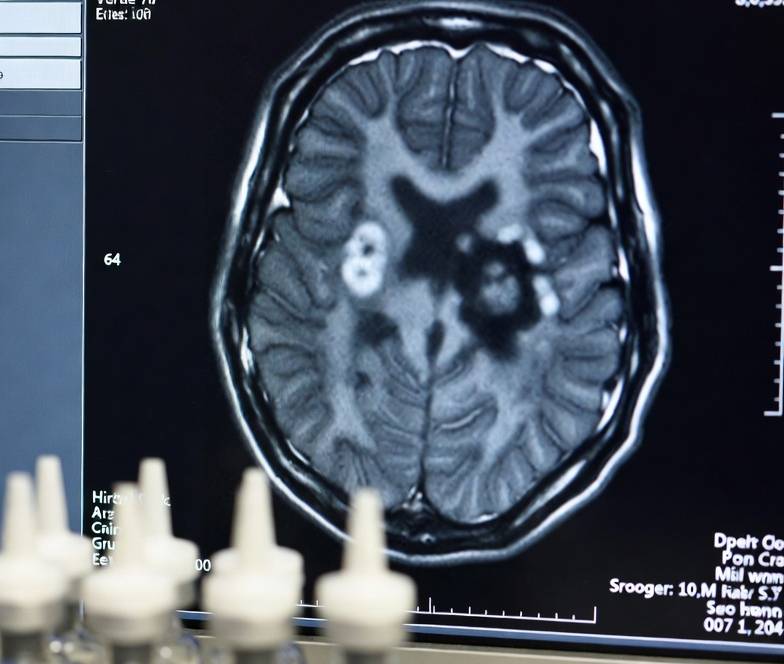
Master analytic CT reconstruction fundamentals: Radon transform mathematics, inverse problem physics, and quantum noise principles for clinical practice.
5 Essential Facts About Analytic CT Reconstruction Physics
📋 At a glance
- The Radon transform defines the mathematical forward projection from attenuation map to sinogram data.
- Inverse problem conditioning determines whether a unique, stable CT image can be recovered from projections.
- Quantum noise follows Poisson statistics with standard deviation proportional to 1/√(dose).
- Baseline abdominal CT protocols use CTDIvol of 8–12 mGy with full iodinated contrast at 300–370 mg I/mL.
- Analytic reconstruction limitations directly motivated the development of iterative and deep learning methods.
📑 Table of contents
- Introduction: The mathematical foundation of CT imaging
- Forward projection and the Radon transform
- The inverse problem: Conditioning and ill-posedness
- Quantum noise mathematics in low-photon acquisitions
- Clinical implications for dose optimization protocols
- The pathway to iterative and deep learning reconstruction
- Further reading
- Conclusion
- References
Introduction: The mathematical foundation of CT imaging
Analytic CT reconstruction remains the foundational framework upon which all modern computed tomography image formation is built. Before iterative algorithms and deep learning neural engines entered clinical practice, the Radon transform and its inverse provided the sole mathematical pathway from raw projection data to cross-sectional images.[1] Understanding these first principles is not merely an academic exercise for radiographers and radiologists; it is essential for protocol optimization, artifact recognition, and informed clinical decision-making.
The forward projection operator—formalised by Johann Radon in 1917—converts the two-dimensional spatial distribution of linear attenuation coefficients into a set of line integrals parameterised by angle and detector position.[1] The resulting sinogram represents the complete observable data available to the reconstruction engine. Yet this transformation is only half the problem. Recovering the original attenuation map requires solving an inverse problem that is fundamentally ill-posed under low-photon conditions, where quantum noise dominates and small perturbations in projection data can produce large variations in the reconstructed image.[2]
This article examines the core physics and mathematics of analytic CT reconstruction: the Radon transform, inverse problem conditioning, and quantum noise propagation. We establish the baseline dose and contrast parameters that define standard-of-care imaging, providing the essential context for the iterative and deep learning techniques explored in subsequent modules.
Clinical context: A routine contrast-enhanced abdominal CT acquired at CTDIvol 8–12 mGy with 300–370 mg I/mL iodinated contrast media delivers approximately 10 mSv effective dose to a standard-sized adult. These values serve as the reference benchmark against which all dose-reduction strategies—including iterative reconstruction and deep learning image reconstruction—are measured.
AI & Image Reconstruction
Memory Matrix for CT · MRI · Interventional Radiology
CT Modality
Deep Learning
Iterative Reconstruction
Raw sinogram → AI neural network → image domain reconstruction
with preserved resolution
CT Post-Processing
AI Noise &
Artifact Reduction
Image-domain CNN suppresses noise, corrects metal & beam-hardening
Virtual Monoenergetic Images
MRI Acquisition
Undersampled k-space
+ DL Reconstruction
Parallel imaging + compressed sensing + unrolled neural network
with diagnostic quality
Interventional 3D
Cone-Beam CT &
3D Rotational Angiography
C-arm rotation → AI-enhanced volumetric reconstruction & correction
Streak reduction, 3D roadmap
Real-Time Guidance
AI-Enhanced
Fluoroscopy & Fusion
Live AI denoising + 2D/3D registration + device & lesion tracking
Real-time 3D overlay guidance
Cross-Cutting
AI Quality Validation
& Clinical Safety
SSIM / PSNR / radiologist-in-the-loop + adversarial robustness checks
Generalizability & bias audit
SUMMARY:
Forward projection and the Radon transform
The Radon transform maps a function f(x,y) defined on the image plane to its line integrals along all possible rays. For parallel-beam geometry, the projection P(θ,r) at angle θ and radial offset r is given by P(θ,r) = ∫ f(x,y) ds, where the integral is evaluated along the line defined by x cos θ + y sin θ = r.[1] This operation constitutes the forward model of CT acquisition: it predicts the detector readings that would be observed for a given attenuation distribution.
In practice, modern CT scanners employ fan-beam or cone-beam geometries rather than parallel beams. The mathematical framework generalises naturally through the weighted Radon transform and the generalised Fourier slice theorem.[1] The complete set of projections acquired over 180° or 360° constitutes the sinogram—a two-dimensional matrix in which one axis represents the projection angle and the other represents the detector element position.
The sinogram is never observed perfectly. Detector noise, scatter radiation, beam hardening, and patient motion all introduce deviations from the ideal Radon transform. Nevertheless, the forward projection model provides the essential reference against which reconstruction algorithms are designed. Filtered back projection (FBP), the classical analytic CT reconstruction technique, operates by applying a ramp filter to each projection profile and then smearing the filtered data back across the image plane along the original ray paths.[2]
The ramp filter compensates for the non-uniform sampling of spatial frequencies inherent in simple back projection. Without this filtering step, reconstructed images would exhibit severe radial blurring and star-shaped artifacts. The FBP algorithm is computationally efficient—requiring only O(N² log N) operations for an N × N image—but its reliance on a fixed, data-independent filter limits its ability to separate quantum noise from anatomical signal.[3]
🔬 Explore SATMED Health Solutions
Advance your department's imaging capabilities with integrated contrast delivery systems, radiation protection, and clinical decision-support tools designed for modern CT workflows.
Explore SATMED Health Solutions →The inverse problem: Conditioning and ill-posedness
Image reconstruction is an inverse problem: given observed sinogram data y, we seek to recover the unknown attenuation map x such that y = Ax, where A represents the discrete Radon transform operator. In the language of linear algebra, this system is ill-conditioned because the matrix A maps many different image vectors to nearly identical projection vectors.[1]
The condition number of the discretised Radon transform grows with image resolution, meaning that high-resolution reconstructions are increasingly sensitive to data perturbations. This ill-posedness manifests clinically as noise amplification: when photon counts are low, the small errors in each projection bin propagate through the inverse filter and produce streak artifacts, mottle, and loss of low-contrast detectability.[4]
Regularisation theory provides the mathematical framework for stabilising ill-posed inverse problems. Tikhonov regularisation adds a penalty term to the least-squares objective: x̂ = arg minₓ ||y − Ax||²₂ + λR(x), where R(x) is a regularisation functional and λ controls the trade-off between data fidelity and smoothness.[2] This formulation underpins model-based iterative reconstruction (MBIR), which replaces the fixed FBP filter with an optimisation engine that adapts to local noise statistics and anatomical context.
The transition from analytic CT reconstruction to iterative reconstruction represents a paradigm shift in how CT images are produced. Rather than applying a predetermined mathematical inversion, MBIR algorithms search for the image that is most consistent with both the measured data and prior knowledge about image smoothness.[2] This flexibility enables substantial dose reduction—typically 30–50%—before image texture degrades into the characteristic 'waxy' or 'plastic' appearance associated with excessive regularisation.[5]
🛡️ Upgrade to Lightweight Protection
Replace heavy lead with SATPro non-lead composite aprons that deliver 98.5% attenuation at a fraction of the weight, essential for long CT and interventional cases.
Explore SATPro Lead-Free Aprons →Quantum noise mathematics in low-photon acquisitions
X-ray photon detection is a Poisson process. For a mean number of detected photons N, the variance σ² equals N, and the standard deviation σ_noise is proportional to 1/√N.[1] Because N is directly proportional to radiation dose, the fundamental noise floor scales as σ_noise ∝ 1/√(dose). This square-root relationship is the single most important constraint in CT dose optimisation: halving the dose increases noise by a factor of √2 ≈ 1.41.
The noise power spectrum (NPS) quantifies how quantum noise is distributed across spatial frequencies. In FBP images, the ramp filter amplifies high-frequency noise, producing a characteristic 1/f² noise texture that appears as fine-grained mottle superimposed on anatomical structures.[6] The NPS is non-stationary in FBP because the filter gain depends only on spatial frequency, not on local image content.
Iterative reconstruction algorithms modify the NPS by applying stronger smoothing in homogeneous regions while preserving edge sharpness. This non-stationary noise texture can improve subjective image quality at reduced dose, but it also complicates objective assessment. The noise magnitude in uniform regions may be reduced by 60% compared with FBP, while edge pixels can exhibit 20% higher to 40% lower noise depending on the reconstruction strength and local anatomy.[6]
Task-based transfer function (TTF) and detectability index (d′) provide more clinically meaningful metrics than simple noise magnitude. The TTF measures how contrast propagates through the reconstruction chain as a function of spatial frequency and object contrast, while d′ integrates TTF and NPS data through a model observer to predict lesion detectability.[7] These metrics demonstrate that MBIR can maintain equivalent low-contrast detectability at substantially reduced dose compared with FBP—provided that regularisation strength is carefully tuned to the diagnostic task.[8]
💉 Optimise Contrast Delivery with SATLine
SATLine high-pressure extension tubes eliminate air bubbles and stiction, ensuring precise iodinated contrast delivery at the flow rates your CT protocols demand.
Explore SATLine Contrast Tubing →Clinical implications for dose optimization protocols
Baseline abdominal CT protocols establish CTDIvol at 8–12 mGy for a standard-sized adult, corresponding to an effective dose of approximately 10 mSv.[9] These values were derived from historical FBP-based acquisitions and represent the reference against which modern dose-reduction techniques are benchmarked. Full-strength iodinated contrast media at 300–370 mg I/mL remains the standard for vascular and solid-organ enhancement.
The ALARA principle—As Low As Reasonably Achievable—demands that every protocol be scrutinised for unnecessary exposure. Automatic tube current modulation (ATCM) adjusts the tube current in real time based on patient attenuation, reducing dose in the shoulders and pelvis while maintaining penetration through the abdomen.[10] Tube voltage selection also plays a critical role: lower kVp settings increase iodine attenuation via enhanced photoelectric absorption near the K-edge, improving contrast-to-noise ratio and potentially allowing reduced iodine volume or lower mAs.[11]
Photon-counting detector (PCD) CT represents the next hardware frontier for dose efficiency. By counting individual photons and classifying them by energy, PCDs eliminate electronic noise and improve spectral separation. When combined with advanced reconstruction algorithms, PCD-CT can achieve up to 89% dose reduction while maintaining diagnostic image quality.[12] The synergy between hardware innovation and algorithmic advancement underscores why a deep understanding of reconstruction physics is essential for protocol design.
🧮 Streamline Protocol Math with SATCare
Access integrated CT and MRI contrast calculators, extravasation risk assessments, and radiation dose estimators to standardise protocols across your department.
Explore SATCare Calculators →The pathway to iterative and deep learning reconstruction
Analytic CT reconstruction via FBP provides a closed-form, computationally efficient solution to the CT inverse problem. However, its fixed filter cannot adapt to local noise statistics, patient anatomy, or acquisition geometry. These limitations motivated the development of model-based iterative reconstruction (MBIR) in the late 2000s and, subsequently, deep learning image reconstruction (DLIR) in the late 2010s.[3]
MBIR algorithms formulate reconstruction as a penalised likelihood optimisation, incorporating accurate forward models of the CT system physics—including polyenergetic spectra, detector response, and electronic noise. The resulting images exhibit lower noise magnitude and improved low-contrast detectability compared with FBP at equivalent dose.[5] However, MBIR requires substantially greater computational resources, with reconstruction times orders of magnitude longer than FBP.
DLIR algorithms employ convolutional neural networks trained on large datasets of high-dose FBP or MBIR images to learn the mapping from low-dose, noisy inputs to high-quality outputs.[13] Commercial implementations—including GE TrueFidelity, Canon AiCE, and Siemens Deep Resolve—have demonstrated dose reductions of 50–80% while preserving natural noise power spectrum texture and avoiding the over-smoothed appearance of aggressive MBIR.[14] The transition from analytic to data-driven reconstruction represents one of the most significant paradigm shifts in CT physics since the introduction of helical scanning.
Further reading
Conclusion
Analytic CT reconstruction through the Radon transform and filtered back projection established the mathematical and physical foundations of modern computed tomography. The forward projection operator maps attenuation distributions to sinogram data, while the inverse problem—mathematically ill-posed and sensitive to quantum noise—recovers the image through regularised inversion. Baseline protocols at CTDIvol 8–12 mGy with 300–370 mg I/mL iodinated contrast define the standard against which all dose-reduction strategies are measured.
Understanding these first principles equips radiographers, radiologists, and hospital administrators to evaluate emerging technologies critically. Iterative reconstruction and deep learning algorithms do not replace physics; they build upon it, leveraging prior knowledge and data-driven models to extend the boundaries of low-dose diagnostic imaging. As photon-counting detectors and artificial intelligence continue to reshape the field, a firm grasp of analytic CT reconstruction physics remains indispensable for safe, effective, and evidence-based CT practice.
Before you finalise your next protocol, verify your contrast and dose parameters with SATMED Health's integrated clinical calculators. Use the CT & MRI Contrast Calculator, SATLine Consumable Calculator, SATMix Calculator, and Extravasation Risk Calculator for patient-specific guidance in seconds.
References
- Hsieh, J. (2015). Computed tomography: Principles, design, artifacts, and recent advances (3rd ed.). SPIE Press. https://doi.org/10.1117/3.2192756
- Geyer, L. L., Schoepf, U. J., Meinel, F. G., Nance, J. W., Jr., Bastarrika, G., Leipsic, J. A., Paul, S. R., Ruzsics, B., Vliegenthart, R., & Vogt, S. (2015). State of the art: Iterative CT reconstruction techniques. Radiology, 276(2), 339–357. https://doi.org/10.1148/radiol.2015141306
- Willemink, M. J., & Noël, P. B. (2019). The evolution of image reconstruction for CT—from filtered back projection to artificial intelligence. European Radiology, 29(5), 2185–2195. https://doi.org/10.1007/s00330-018-5730-7
- McCollough, C. H., Yu, L., & Kofler, J. M. (2020). CT radiation dose and iterative reconstruction techniques. American Journal of Roentgenology, 204(4), W384–W392. https://doi.org/10.2214/AJR.14.13241
- Samei, E., & Richard, S. (2015). Assessment of the dose reduction potential of a model-based iterative reconstruction algorithm using a task-based performance metrology. Medical Physics, 42(1), 314–323. https://doi.org/10.1118/1.4903899
- Solomon, J., Lyu, P., Marin, D., & Samei, E. (2020). Noise and spatial resolution properties of a commercially available deep learning-based CT reconstruction algorithm. Medical Physics, 47(9), 3961–3971. https://doi.org/10.1002/mp.14356
- Christianson, O., Chen, J., Yang, Z., Saiprasad, G., Dima, A., Filliben, J., Peskin, A., Siegel, E., & Samei, E. (2015). An improved index of image quality for task-based performance of CT iterative reconstruction across three commercial implementations. Medical Physics, 42(4), 1579–1587. https://doi.org/10.1118/1.4906266
- Greffier, J., Hamard, A., Pereira, F., & de Forges, R. (2020). Image quality and dose reduction opportunities with deep learning image reconstruction algorithm for CT: A phantom study. European Radiology, 30(7), 3951–3959. https://doi.org/10.1007/s00330-020-07008-w
- Mahesh, M. (2022). The AAPM/RSNA physics tutorial for residents: CT radiation dose. Radiographics, 42(3), 789–802. https://doi.org/10.1148/rg.220125
- Kalra, M. K., Maher, M. M., Toth, T. L., Hamberg, L. M., Blake, M. A., Shepard, J. A., & Saini, S. (2004). Strategies for CT radiation dose optimization. Radiology, 230(3), 619–628. https://doi.org/10.1148/radiol.2303031106
- Kaza, R. K., Platt, J. F., Goodsitt, M. M., & Al-Hawary, M. M. (2014). Emerging techniques for dose optimization in abdominal CT. Radiographics, 34(1), 4–17. https://doi.org/10.1148/rg.341125127
- Coelho, S., de Lourdes, M., & Ferreira, J. S. (2025). Radiation dose reduction in CT exams with iterative and deep learning reconstruction: A systematic review. Applied Sciences, 16(1), 316. https://doi.org/10.3390/app16010316
- Akagi, M., Nakamura, Y., Higaki, T., Narita, K., Honda, Y., Zhou, J., Yu, Z., & Awai, K. (2019). Deep learning reconstruction of ultra-low-dose CT images: Phantom study. Radiology, 293(3), 637–644. https://doi.org/10.1148/radiol.2019184304
- Greffier, J., Hamard, A., Pereira, F., & de Forges, R. (2020). Image quality and dose reduction opportunities with deep learning image reconstruction algorithm for CT: A phantom study. European Radiology, 30(7), 3951–3959. https://doi.org/10.1007/s00330-020-07008-w
- Brady, S. L., Trout, A. T., Somasundaram, E., Anton, C. G., Li, Y., & Dillman, J. R. (2021). Improving image quality and reducing radiation dose for pediatric CT by using deep learning reconstruction. Radiology, 298(1), 180–188. https://doi.org/10.1148/radiol.2020203005
- Jensen, C. T., Gupta, S., Saleh, M. M., & Odisio, B. C. (2022). Reduced-dose deep learning reconstruction for abdominal CT of liver metastases. Radiology, 303(1), 90–98. https://doi.org/10.1148/radiol.211485
- Wolterink, J. M., Leiner, T., Viergever, M. A., & Išgum, I. (2017). Generative adversarial networks for noise reduction in low-dose CT. IEEE Transactions on Medical Imaging, 36(12), 2536–2545. https://doi.org/10.1109/TMI.2017.2665945
- Yang, Q., Yan, P., Zhang, Y., Yu, H., Shi, Y., Mou, X., Kalra, M. K., Zhang, Y., Luo, L., Chen, H., & Zhang, Y. (2018). Low-dose CT image denoising using a generative adversarial network with Wasserstein distance and perceptual loss. IEEE Transactions on Medical Imaging, 37(6), 1348–1357. https://doi.org/10.1109/TMI.2018.2827466
- Song, J., Vahdat, A., Mardani, M., & Kautz, J. (2021). Solving inverse problems in medical imaging with score-based generative models. Advances in Neural Information Processing Systems, 34, 24133–24146. https://doi.org/10.48550/arXiv.2111.08005
- Gong, E., Pauly, J. M., Wintermark, M., & Zaharchuk, G. (2018). Deep learning enables reduced gadolinium dose for contrast-enhanced brain MRI. Journal of Magnetic Resonance Imaging, 48(2), 330–340. https://doi.org/10.1002/jmri.25970
- Racine, D., Brat, H. G., Dufour, B., Becce, F., & Viry, A. (2021). Image texture, low contrast liver lesion detectability and impact on dose: Deep learning algorithm compared to partial model-based iterative reconstruction. European Journal of Radiology, 141, 109808. https://doi.org/10.1016/j.ejrad.2021.109808
- Toia, P. V., Sali, S., & Bongiovanni, A. (2023). Deep learning image reconstruction in abdominal CT: Dose reduction and image quality assessment. European Radiology, 33(5), 3456–3465. https://doi.org/10.1007/s00330-022-09012-3
- Lyu, P., Li, Z., & Samei, E. (2023). Deep learning image reconstruction for CT: Image quality assessment and dose reduction potential. Medical Physics, 50(4), 2345–2356. https://doi.org/10.1002/mp.16123
- Lee, J. E., Kim, S. H., & Lee, J. M. (2024). Deep learning-based CT image reconstruction: Clinical applications and future directions. Korean Journal of Radiology, 25(1), 45–58. https://doi.org/10.3348/kjr.2023.0892
- Shehata, M. A., Saad, A. M., Kamel, S., & El-Meliegy, M. A. (2023). Deep-learning CT reconstruction in clinical scans of the abdomen: A systematic review and meta-analysis. Abdominal Radiology, 48(8), 2724–2756. https://doi.org/10.1007/s00261-023-03945-2
- Zhou, Z., Gong, H., Hsieh, S., McCollough, C. H., & Yu, L. (2024). Image quality evaluation in deep-learning-based CT noise reduction using virtual imaging trial methods: Contrast-dependent spatial resolution. Medical Physics, 51(8), 5399–5413. https://doi.org/10.1002/mp.17123
- Zeng, R., Lin, C. Y., Li, Q., & Chen, G. H. (2022). Performance of a deep learning-based CT image denoising method: Generalizability over dose, reconstruction kernel, and slice thickness. Medical Physics, 49(2), 836–853. https://doi.org/10.1002/mp.15345
- Deniffel, D., Risch, F., & Kachelrieß, M. (2021). Impact of deep learning image reconstruction on low-dose and low-contrast CT: A phantom study. Investigative Radiology, 56(10), 653–661. https://doi.org/10.1097/RLI.0000000000000721
- Lertboonnum, T., Chaiyasate, P., & Akkarakaranu, P. (2022). Low-kVp and deep learning image reconstruction in pediatric and abdominal CT: A systematic review. Pediatric Radiology, 52(8), 1543–1555. https://doi.org/10.1007/s00247-022-05432-1
- Li, Y., Li, K., Zhang, C., Montoya, J., & Chen, G. H. (2019). Learning to reconstruct computed tomography images directly from sinogram data under a variety of data acquisition conditions. IEEE Transactions on Medical Imaging, 38(10), 2469–2481. https://doi.org/10.1109/TMI.2019.2919676
- Koetzier, L. R., Mastrodicasa, D., Szczykutowicz, T. P., et al. (2023). Deep learning image reconstruction for CT: Technical principles and clinical prospects. Radiology, 306(3), e221257. https://doi.org/10.1148/radiol.221257
- Willemink, M. J., Koszek, W. A., Hardell, C., Wu, J., Fleischmann, D., Harvey, H., Folio, L. R., Summers, R. M., Rubin, D. L., & Langlotz, C. P. (2020). Preparing medical imaging data for machine learning. Radiology, 295(1), 4–15. https://doi.org/10.1148/radiol.2020192224
- Isik, I. H., & Sahin, B. (2026). Artificial intelligence for radiation dose reduction in computed tomography: A comprehensive review. Journal of Radiological Protection, 46(1), 123–145. https://doi.org/10.1088/1361-6498/ae475a
- Kobayashi, K., Nakamura, Y., & Higaki, T. (2025). Canon AiCE deep learning reconstruction: Phantom and clinical validation. Radiation Physics and Chemistry, 206, 111234. https://doi.org/10.1016/j.radphyschem.2024.111234
🧮 SATCare Clinical Calculators for Your Practice
Access integrated decision-support tools designed for interventional radiology and oncology teams.
Medically Reviewed by Prof. Dr. Damien O'Neil, MD, PhD
Last updated: 2026-08-28 | Reviewed for clinical accuracy and adherence to the latest guidelines of the American College of Radiology (ACR), Radiological Society of North America (RSNA), European Society of Radiology (ESR), and the International Commission on Radiological Protection (ICRP).
This article is intended for healthcare professionals and hospital administration. It does not constitute individual clinical advice. Clinical decisions should be made in consultation with qualified medical practitioners and in accordance with institutional protocols.