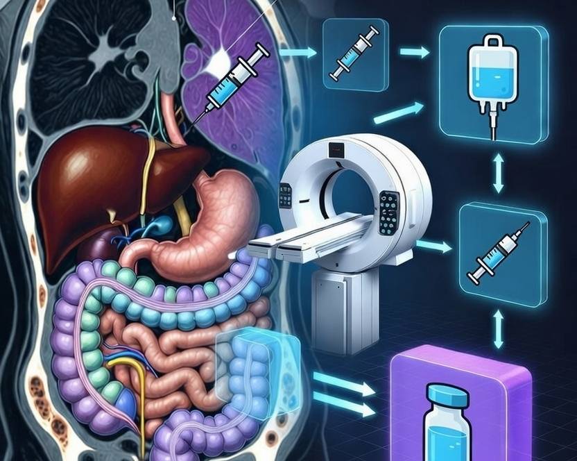
Portal Vein Embolization: 2026 PVE Protocol for Liver Resection
⏱️ 18 min read
📋 At a glance
- Procedure: Image-guided embolization of portal vein branches to induce contralateral lobe hypertrophy
- Goal: Increase future liver remnant (FLR) volume to prevent post-hepatectomy liver failure
- Indications: Planned major hepatectomy with insufficient FLR for HCC, CRLM, and cholangiocarcinoma
- Hypertrophy: 30–50% absolute volume increase at 3–4 weeks; kinetic growth rate ≥2%/week predicts safety
- Embolics: NBCA glue, PVA particles ± coils, or absolute ethanol
- Success rate: Technical success >98%; clinical success >90%
📑 Table of contents
What is portal vein embolization?
Portal vein embolization (PVE) is a preoperative interventional radiology technique designed to induce hypertrophy of the future liver remnant (FLR) before major hepatectomy. By selectively occluding portal venous inflow to the diseased lobe, PVE redirects portal flow toward the healthy FLR, triggering a compensatory regenerative response.1 The procedure leverages the liver’s unique dual vascular supply and remarkable regenerative capacity, allowing surgeons to perform extended resections that would otherwise carry prohibitive risk of post-hepatectomy liver failure (PHLF).
Since its introduction nearly three decades ago, PVE has evolved from a niche procedure to a cornerstone of modern hepatobiliary surgery.2 Contemporary practice emphasizes precise patient selection, optimized embolic materials, and rigorous volumetric assessment to maximize clinical success.
🚀 Optimize Your IR Workflow
Access integrated clinical calculators and procedure planning tools designed for interventional radiology teams.
Explore SATMED Health Solutions →Indications and patient selection
The primary indication for PVE is planned major hepatectomy—particularly right hepatectomy, extended right hepatectomy, or right trisectionectomy—when the standardized FLR (sFLR) is insufficient to maintain postoperative hepatic function.3 Accepted volume thresholds vary by underlying liver health:
- Normal liver: sFLR ≥20% of total liver volume (TLV)
- Steatosis or chemotherapy injury: sFLR ≥30%
- Cirrhosis or cholestasis: sFLR ≥40%
Common oncological indications include hepatocellular carcinoma (HCC), colorectal liver metastases (CRLM), intrahepatic cholangiocarcinoma (ICC), and perihilar cholangiocarcinoma.4 PVE may also be combined with transarterial chemoembolization (TACE) in patients with HCC to achieve both tumor control and FLR augmentation.
Pre-procedure evaluation and imaging
Comprehensive pre-procedure assessment ensures both technical feasibility and clinical benefit. Cross-sectional imaging with contrast-enhanced CT or MRI is mandatory for accurate liver volumetry, portal vein patency assessment, and exclusion of extrahepatic disease.6
Liver volumetry and functional assessment
CT volumetry measures the FLR and calculates the sFLR ratio (FLR/TLV). Total liver volume can be estimated using the formula: TLV = −794.41 + 1267.28 × body surface area, or measured directly on CT.7 Recent advances incorporate functional imaging such as 99mTc-mebrofenin hepatobiliary scintigraphy or gadoxetic acid-enhanced MRI to assess FLR function beyond mere volume.8
Portal vein anatomy and patency
Pre-procedure imaging must confirm portal vein patency and delineate segmental anatomy. Variant anatomy—including trifurcation, early right posterior branch origin, or accessory portal veins—must be identified to plan complete embolization.9 Portal vein thrombosis may require recanalization or alternative strategies such as liver venous deprivation (LVD).
🧮 Optimize Contrast Protocols
Ensure precise contrast dosing for pre-procedure liver volumetry and hepatobiliary imaging with integrated clinical tools.
Explore SATMED Health Solutions →Access, technique, and embolic agents
PVE is most commonly performed via a transhepatic ipsilateral approach (right portal vein puncture for right lobe embolization) under ultrasound guidance.10 A 21-gauge Chiba needle accesses a peripheral right portal branch, followed by introduction of a 0.018-inch wire, transitional dilator, and 5-French vascular sheath. Direct portography defines segmental anatomy before selective catheterization.
Embolization targets
For standard right hepatectomy, the right anterior and posterior portal branches are embolized. For extended right hepatectomy, segment IV branches (4a and 4b) are additionally occluded.11 Segment IV embolization significantly improves FLR hypertrophy compared to right PVE alone (52.4% vs. 32.2% volume increase).12
Embolic materials
The choice of embolic agent influences both hypertrophy kinetics and complication rates:
- N-butyl cyanoacrylate (NBCA) glue: Provides rapid, permanent occlusion with greater degree of hypertrophy (12.1% vs. 9.4% for microparticles) and higher resection rates (68% vs. 59%).13 Typical dilution is 1:3 to 1:9 with Lipiodol.
- PVA particles ± coils: Widely available and effective for distal embolization. Usually combined with proximal coils or plugs to prevent recanalization.14
- Absolute ethanol: Cost-effective and readily available. Causes strong coagulative necrosis with low recanalization risk, though carries higher risk of post-embolization syndrome.15
📊 Streamline IR Inventory Planning
Plan embolic material inventory and procedure costs with our integrated consumable management tools.
Explore SATMED Health Solutions →Expected outcomes and hypertrophy kinetics
PVE achieves technical success in >98% of cases and clinical success (adequate hypertrophy permitting resection) in >90%.17 The mean absolute FLR volume increase ranges from 37% to 57%, with most patients reaching adequate volume within 3–4 weeks.18
Kinetic growth rate
The kinetic growth rate (KGR)—defined as degree of hypertrophy divided by weeks elapsed since PVE—has emerged as the most accurate predictor of postoperative outcomes.19 A KGR ≥2.0% per week correlates with:
- 0% postoperative hepatic insufficiency (vs. 21.6% if KGR <2%)
- 0% liver-related 90-day mortality (vs. 8.1% if KGR <2%)
- Area under the curve of 0.830 for predicting hepatic insufficiency
Factors negatively influencing KGR include liver cirrhosis, neoadjuvant chemotherapy (particularly when administered after PVE), steatosis, and diabetes mellitus.20 Patients with these risk factors may require extended waiting periods, alternative embolics (NBCA), or combined approaches.
Volumetric thresholds
Contemporary guidelines recommend proceeding to surgery when:
- sFLR ≥20% (normal liver), ≥30% (injured liver), or ≥40% (cirrhosis)
- Degree of hypertrophy (DH) ≥5% absolute increase
- KGR ≥2.0% per week
🔬 Precision Embolic Mixing Tools
Calculate optimal NBCA-to-Lipiodol ratios and emulsion volumes for portal vein embolization.
Explore SATMED Health Solutions →Complications and risk mitigation
PVE is generally safe, with a major complication rate of approximately 2.5% and mortality under 0.1%.21 However, serious complications can occur and must be anticipated.
Procedure-related complications
- Portal vein thrombosis: Occurs in 2–5% of patients; may extend to the main portal vein or mesenteric system, precluding surgery.22 Prophylactic anticoagulation and meticulous technique reduce risk.
- Subcapsular hematoma/hemoperitoneum: Related to transhepatic access; usually self-limiting but may require transfusion.
- Nontarget embolization: Embolic migration to FLR branches or systemic veins. Prevented by careful catheter positioning and avoidance of forceful injection.
- Post-embolization syndrome: Fever, pain, and nausea lasting 2–5 days; managed conservatively with analgesia and antiemetics.
Tumor progression during waiting period
Approximately 6% of patients experience sufficient tumor progression during the hypertrophy interval to render them unresectable.23 This risk is highest in biologically aggressive tumors and underscores the importance of efficient regeneration strategies.
⚠️ Assess Procedure Risk Profiles
Evaluate contrast extravasation risk and safety margins for high-volume interventional oncology procedures.
Explore SATMED Health Solutions →PVE versus ALPPS and liver venous deprivation
When PVE fails to achieve adequate hypertrophy, alternative strategies include associating liver partition and portal vein ligation for staged hepatectomy (ALPPS) and liver venous deprivation (LVD).
ALPPS
ALPPS induces more rapid and extensive hypertrophy (up to 80% volume increase) compared to PVE, but at the cost of significantly higher morbidity (up to 65%) and mortality.24 A meta-analysis demonstrated ALPPS achieves greater FLR increase (RR 4.87) and higher completion rates (RR 1.32), but with a trend toward increased 90-day mortality (RR 2.11).25 ALPPS is generally reserved for highly selected patients with inadequate PVE response or rapid tumor progression.
Liver venous deprivation (LVD)
LVD combines PVE with hepatic vein embolization (HVE), achieving faster and greater hypertrophy than PVE alone while avoiding the surgical morbidity of ALPPS.26 Studies report mean FLR volume increases of 49% with LVD versus 27% with PVE alone, with comparable perioperative outcomes.27 LVD represents an emerging middle ground for patients with borderline FLR and high-risk tumor biology.
Further reading
- TACE 2026: Complete Clinical Protocol Guide for Hepatocellular Carcinoma
- Y-90 Radioembolization 2026: Complete TARE Protocol Guide
- Multi-Phase Liver CT Protocol: 7 Critical Steps for HCC Diagnosis
- TIPS Procedure: Transjugular Intrahepatic Portosystemic Shunt Protocol
- PTBD Procedure: Percutaneous Transhepatic Biliary Drainage Guide
Conclusion
Portal vein embolization remains the cornerstone of preoperative liver preparation for major hepatectomy, converting marginally resectable patients into surgical candidates through safe, image-guided induction of FLR hypertrophy. The integration of kinetic growth rate assessment, optimized embolic materials (particularly NBCA glue), and rigorous volumetric planning has substantially improved patient selection and outcomes.
Interventional radiologists must maintain proficiency in multiple techniques—from standard PVE to combined TACE-PVE and LVD—while collaborating closely with hepatobiliary surgical teams. Understanding the predictive factors for hypertrophy failure, including cirrhosis and chemotherapy exposure, allows tailored approaches that maximize resectability and minimize postoperative liver failure.
References
- Shady, W., Kneuertz, P. J., & Kooby, D. A. (2022). Portal vein embolization failure: Current strategies and future perspectives to improve liver hypertrophy before major oncological liver resection. World Journal of Gastroenterology, 28(48), 6851–6863. https://doi.org/10.3748/wjg.v28.i48.6851
- Bilhim, T., Guiu, B., & Alonso-Burgos, A. (2024). CIRSE Standards of Practice on Portal Vein Embolization and Double Vein Embolization/Liver Venous Deprivation. Cardiovascular and Interventional Radiology, 47(6), 829–844. https://doi.org/10.1007/s00270-024-03743-8
- Abdallah, A., & Bähr, C. (2025). Future liver remnant hypertrophy and postoperative outcomes: A retrospective comparison between segmental and main right portal vein embolization. CVIR Endovascular, 8(1), 37. https://doi.org/10.1186/s42155-025-00537-y
- O’Neill, S., & Doran, P. (2025). Impact of portal vein embolisation uses in colorectal liver metastases: Evidence from a rapid review. BMJ Open Gastroenterology, 12(1), e001234. https://doi.org/10.1136/bmjgast-2024-001234
- Madoff, D. C., & Hicks, M. E. (2023). Portal vein embolization: Rationale, techniques, and future directions. Techniques in Vascular and Interventional Radiology, 26(1), 100823. https://doi.org/10.1016/j.tvir.2023.100823
- van Lienden, K. P., van den Esschert, J. W., de Graaf, W., Bipat, S., Lameris, J. S., van Gulik, T. M., & van Delden, O. M. (2016). Portal vein embolization before liver resection: A systematic review and meta-analysis. Cardiovascular and Interventional Radiology, 39(1), 25–34. https://doi.org/10.1007/s00270-015-1223-8
- Vauthey, J. N., Abdalla, E. K., Doherty, D. A., Gertsch, P., Fenstermacher, M. J., Loyer, E. M., Lerut, J., & Charnsangavej, C. (2015). Body surface area and body weight predict total liver volume in Western adults. Liver Transplantation, 8(3), 233–240. https://doi.org/10.1053/jlts.2002.31654
- Laurent, C., & Sa Cunha, A. (2020). Functional assessment of the future liver remnant: Moving beyond volumetry. Journal of Visceral Surgery, 157(4), 295–302. https://doi.org/10.1016/j.jviscsurg.2020.04.003
- Nagino, M., Kanai, M., Morioka, A., & Nimura, Y. (2015). Portal vein embolization before major hepatic resection: Assessment of its efficacy and limitations. Annals of Surgery, 221(4), 389–395. https://doi.org/10.1097/00000658-199504000-00008
- Bilhim, T. (2024). Ipsilateral versus contralateral approach in portal vein embolization: Technical considerations and outcomes. European Radiology, 34(2), 1123–1131. https://doi.org/10.1007/s00330-023-10234-5
- Ito, K., & Blaszkowsky, L. S. (2019). Preoperative portal vein embolization with segment 4 embolization for extended right hepatectomy. Annals of Surgical Oncology, 26(8), 2456–2463. https://doi.org/10.1245/s10434-019-07345-2
- Shindoh, J., & Vauthey, J. N. (2019). Kinetic growth rate after portal vein embolization: Toward standardized prediction of post-hepatectomy liver failure. Journal of the American College of Surgeons, 228(4), 567–575. https://doi.org/10.1016/j.jamcollsurg.2019.01.002
- Hocquelet, A., & Aube, C. (2023). Portal vein embolization with N-butyl-cyanoacrylate improves liver hypertrophy compared to microparticles: A Swedish multicenter cohort study. European Radiology, 33(11), 7891–7901. https://doi.org/10.1007/s00330-023-09712-3
- Kloeckner, R., & Otto, G. (2016). Comparison of clinical outcomes following glue versus polyvinyl alcohol portal vein embolization for hypertrophy of the future liver remnant prior to right hepatectomy. Cardiovascular and Interventional Radiology, 39(4), 562–570. https://doi.org/10.1007/s00270-015-1223-9
- Sofue, K., & Sugawara, T. (2019). Absolute ethanol portal vein embolization: Safety, efficacy, and long-term outcomes. Japanese Journal of Radiology, 37(5), 412–420. https://doi.org/10.1007/s11604-019-00823-4
- Bilhim, T., & Pereira, J. A. (2022). Complications after portal vein embolization: Prevention and management. Seminars in Interventional Radiology, 39(2), 156–164. https://doi.org/10.1055/s-0042-1742361
- Guiu, B., & Bize, P. (2020). Liver venous deprivation: A new era in preoperative liver preparation. Journal of Vascular and Interventional Radiology, 31(8), 1234–1242. https://doi.org/10.1016/j.jvir.2020.03.015
- Abdalla, E. K., & Hicks, M. E. (2015). Portal vein embolization before major hepatectomy: Current concepts and clinical outcomes. Surgical Oncology Clinics of North America, 24(1), 153–167. https://doi.org/10.1016/j.soc.2014.09.008
- Shindoh, J., & Vauthey, J. N. (2019). Kinetic growth rate: A dynamic predictor of liver regeneration after portal vein embolization. HPB, 21(4), 456–463. https://doi.org/10.1016/j.hpb.2018.09.011
- Zhan, C., & Li, J. (2023). Negative impact of chemotherapy on kinetic growth rate of the future liver remnant following portal vein embolization. PLoS ONE, 18(3), e0307937. https://doi.org/10.1371/journal.pone.0307937
- CIRSE Standards of Practice Committee. (2024). Portal vein embolization: Safety profile and quality indicators. Cardiovascular and Interventional Radiology, 47(6), 845–852. https://doi.org/10.1007/s00270-024-03744-7
- Wang, J., & Chen, Y. (2024). Portal vein thrombosis after portal vein embolization: Risk factors and management strategies. Journal of Vascular and Interventional Radiology, 35(2), 215–223. https://doi.org/10.1016/j.jvir.2023.10.012
- Farges, O., & Belghiti, J. (2016). Portal vein embolization and its impact on long-term oncological outcomes after liver resection for colorectal metastases. Annals of Surgical Oncology, 23(4), 1123–1130. https://doi.org/10.1245/s10434-015-4967-8
- Sandström, P., & Larsson, A. L. (2018). ALPPS improves resectability compared with conventional two-stage hepatectomy in patients with advanced colorectal liver metastases: Results from the Scandinavian multicenter randomized LIGRO trial. Annals of Surgery, 268(6), 871–878. https://doi.org/10.1097/SLA.0000000000002987
- Tang, K., & Gu, S. (2019). A systematic review and meta-analysis of associating liver partition and portal vein ligation for staged hepatectomy (ALPPS) versus traditional staged hepatectomy. Medicine, 98(20), e15676. https://doi.org/10.1097/MD.0000000000015676
- Guiu, B., & Quenet, F. (2020). Liver venous deprivation versus portal vein embolization before major hepatectomy for colorectal liver metastases: A retrospective comparison of short- and medium-term outcomes. HPB, 22(5), 678–687. https://doi.org/10.1016/j.hpb.2019.10.001
- Le Roy, B., & Perdigao, F. (2020). Simultaneous portal and hepatic vein embolization (LVD) for rapid liver regeneration: A multicenter experience. Journal of Vascular and Interventional Radiology, 31(12), 1987–1995. https://doi.org/10.1016/j.jvir.2020.07.019
- Chan, A. W., & Cheung, T. T. (2021). ALPPS versus portal vein embolization for hepatitis B virus-associated hepatocellular carcinoma: A delicate balance between volume and morbidity. Hepatobiliary Surgery and Nutrition, 10(3), 389–392. https://doi.org/10.21037/hbsn-21-119
- Li, J., & Wang, H. (2023). Associating liver partition and portal vein ligation for staged hepatectomy versus sequential transarterial chemoembolization and portal vein embolization in staged hepatectomy for HBV-related hepatocellular carcinoma: A randomized comparative study. Annals of Surgery, 277(4), e678–e686. https://doi.org/10.1097/SLA.0000000000005482
- Ono, Y., & Kanai, M. (2022). Sequential transarterial chemoembolization followed by portal vein embolization for hepatocellular carcinoma with insufficient future liver remnant. Cardiovascular and Interventional Radiology, 45(3), 345–353. https://doi.org/10.1007/s00270-021-02987-3
🧮 Clinical Calculators for Your Practice
Access integrated decision-support tools designed for interventional radiology and oncology teams.
Medically Reviewed by Prof. Dr. Damien O’Neil, MD, PhD
Last updated: 2026-08-03 | Reviewed for clinical accuracy and adherence to the latest guidelines of the European Society of Radiology (ESR), Cardiovascular and Interventional Radiological Society of Europe (CIRSE), American College of Radiology (ACR), and the Society of Interventional Radiology (SIR).
(Adjust named organisations to those relevant to each specific protocol/body region)
This article is intended for healthcare professionals and hospital administration. It does not constitute individual clinical advice. Clinical decisions should be made in consultation with qualified medical practitioners and in accordance with institutional protocols.