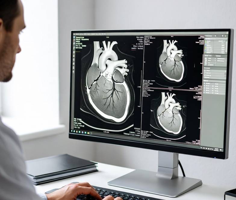
Master the female pelvis MRI protocol with a step-by-step framework covering high-resolution sagittal and oblique-axial T2, dual axial T1 with and without fat saturation, bladder-motion artifact control, and the scanning, interpretive, and clinical pitfalls that most often undermine accurate uterine and ovarian characterization.
Female Pelvis MRI Protocol: The Complete Radiographer & Radiologist Guide
🧲 Sequences Used
- High-resolution sagittal T2 (uterus/cervix long axis)
- Oblique-axial T2 perpendicular to the endometrial canal/cervical canal
- Axial T1 with and without fat saturation
- Axial DWI (adnexal/endometrial lesion characterization)
💉 Contrast Protocol
10–15 mL (0.1 mmol/kg) gadolinium-based agent at 2.0 mL/s, followed by a 100 mL saline chaser at 2.0 mL/s, when dynamic post-contrast assessment of an adnexal or endometrial lesion is clinically indicated.
🎯 Artifact Reduction
Primary artifact: bladder motion/pulsation. Remedy: instruct partial bladder filling before scanning and apply anterior abdominal wall saturation bands to suppress ghosting propagating across the pelvis.
⚠️ Key Pitfalls
- Radiographers: scanning with an overdistended or empty bladder
- Radiologists: bladder pulsation ghost mistaken for an adnexal lesion
- Referrers: acting on ultrasound and MRI reports as directly interchangeable
- Introduction
- Female Pelvic Anatomy Essentials
- MR Tissue Relaxation Values
- Scanning Technique — 10 Steps
- Contrast Media Protocol
- Specific Absorption Rate & Dose Reduction
- Top 10 Pathologies
- Pitfalls — Radiographers
- Pitfalls — Radiologists
- Pitfalls — Non-Radiology Physicians
- Pitfall Comparison Summary
- AI & Automation in Female Pelvis MRI
- Further Reading
- Reducing Artefacts with Patients and Parameters
- Parallel Imaging Protocols and Parameters
- Conclusion
- References
Introduction
A well-executed female pelvis MRI protocol is the reference-standard problem-solving tool for uterine and ovarian pathology that ultrasound leaves equivocal, and the definitive local staging modality for endometrial and cervical carcinoma. Because pelvic organ position, orientation, and even zonal anatomy shift meaningfully with bladder volume and bowel peristalsis, this protocol is unusually dependent on deliberate patient preparation — a detail that separates a diagnostic study from one degraded by exactly the motion artifact this guide is built around.
Unlike many oncologic MRI protocols where contrast is mandatory, female pelvis MRI is frequently performed non-contrast for benign indications such as fibroid mapping or Müllerian anomaly characterization, with dynamic post-contrast imaging reserved for endometrial/cervical cancer staging and indeterminate adnexal mass characterization. Knowing which clinical question is being asked therefore directly shapes whether contrast is needed at all — a protocol decision this guide addresses explicitly in the contrast section below.
This guide walks through the complete female pelvis MRI workflow: the uterine and ovarian anatomy that dictates sequence planning, relevant relaxation values, a ten-step scanning technique, the contrast protocol and when it is and is not required, SAR-conscious parameter selection, the top ten pathologies the protocol is built to detect, and the distinct pitfalls that affect radiographers at the console, radiologists at the workstation, and referring gynecologists acting on the report.
Reliable Contrast Delivery with SATline
Consistent, kink-resistant tubing supports reproducible dynamic timing when adnexal or endometrial contrast assessment is indicated.
Female Pelvic Anatomy Essentials
The uterus, cervix, and ovaries occupy the true pelvis between the bladder anteriorly and the rectosigmoid colon posteriorly — a sandwich relationship that makes bladder and bowel preparation genuinely inseparable from image quality, not merely a courtesy to patient comfort.
Uterine zonal anatomy
On T2-weighted imaging, the normal uterine corpus displays three distinct signal layers. The endometrium is markedly T2 hyperintense, varying in thickness with menstrual cycle phase. The junctional zone — the innermost myometrial layer — is a thin, well-defined band of relatively low T2 signal, and its thickness is the central discriminator in adenomyosis diagnosis (a junctional zone ≥12 mm is considered abnormal). The outer myometrium is intermediate T2 signal, relatively homogeneous in the normal uterus, and the layer against which leiomyoma and myometrial tumor invasion are assessed.
Cervix
The cervix shows a comparable, though distinct, zonal pattern: a central T2-hyperintense mucosal/endocervical canal signal, surrounded by a low-signal-intensity fibrous cervical stroma — the cervical stromal ring — whose integrity is the key determinant of parametrial invasion in cervical cancer staging.
Ovaries and adnexa
The ovaries are variably positioned but most commonly located in the ovarian fossa lateral to the uterus, and their MRI appearance varies substantially with menstrual cycle phase due to follicular development and corpus luteum formation. The adnexa — ovaries, fallopian tubes, and surrounding broad ligament tissue — is the site of origin of the mass lesions this protocol is frequently obtained to characterize, and normal fallopian tubes are not typically visualized unless distended by fluid, blood, or pus.
MR Tissue Relaxation Values
Understanding baseline T1 and T2 relaxation times of normal uterine and ovarian tissue underpins correct recognition of the zonal and adnexal signal abnormalities this protocol is designed to detect.
| Structure | T1 (ms) @ 1.5T | T1 (ms) @ 3T | T2 (ms) @ 1.5T | T2 (ms) @ 3T |
|---|---|---|---|---|
| Endometrium (secretory phase) | ~1300–1500 | ~1600–1850 | ~110–140 | ~95–120 |
| Junctional zone | ~900–1000 | ~1150–1300 | ~35–50 | ~30–42 |
| Outer myometrium | ~1150–1250 | ~1450–1550 | ~70–90 | ~60–78 |
| Cervical stroma | ~950–1050 | ~1200–1350 | ~40–55 | ~35–48 |
| Ovarian follicular fluid | ~2200–2800 | ~2700–3200 | >150 | >150 |
| Skeletal muscle (reference) | ~870 | ~1420 | ~47 | ~32 |
This relaxation profile explains the fundamental “black stripe” appearance of the junctional zone relative to the brighter outer myometrium and endometrium, and why loss or thickening of this low-signal band — rather than myometrial signal change alone — is the primary MRI signature of adenomyosis. It also explains why simple ovarian follicular cysts appear uniformly hyperintense on T2, similar to simple cysts elsewhere in the body, while a hemorrhagic cyst or endometrioma shows the markedly different T1-hyperintense, T2 “shading” pattern discussed in the pathology section below.
Scanning Technique — 10 Steps
- Patient preparation and bladder instruction. Instruct the patient to partially empty the bladder 1–2 hours before scanning, aiming for a moderately, not maximally, distended bladder — sufficient to displace bowel loops without inducing motion-provoking discomfort or urgency.
- Antispasmodic/bowel prep consideration. Consider an antispasmodic agent to reduce bowel peristalsis in patients with a history of motion-degraded prior studies, per departmental protocol.
- Coil selection and positioning. Use a torso/pelvic phased-array coil positioned to cover the uterine fundus superiorly and the full cervix and upper vagina inferiorly.
- Sagittal T2 localizer. Acquire a high-resolution sagittal T2 sequence first — this defines the uterine and cervical long axis used to prescribe every subsequent oblique plane.
- Oblique-axial T2 (short axis of the uterine corpus). Prescribe perpendicular to the endometrial canal as visualized on the sagittal localizer, essential for accurate junctional zone and myometrial invasion assessment.
- Oblique-axial T2 (short axis of the cervix), when relevant. Prescribe perpendicular to the cervical canal for cervical stromal ring assessment in suspected or known cervical pathology.
- Axial T1 without fat saturation. Acquire a standard axial T1 sequence across the full pelvis to identify T1-hyperintense lesions such as hemorrhagic cysts, endometriomas, or dermoid cysts.
- Axial T1 with fat saturation. Acquire the paired fat-saturated sequence to confirm macroscopic fat (dermoid) versus blood products (endometrioma/hemorrhagic cyst) — fat suppresses, blood does not.
- Axial DWI. Acquire diffusion-weighted imaging with ADC map generation to support adnexal and endometrial lesion characterization, particularly for suspected malignancy.
- Dynamic post-contrast 3D T1 fat-saturated (when indicated). For endometrial/cervical cancer staging or indeterminate adnexal masses, administer the gadolinium bolus and acquire a dynamic series to assess myometrial invasion depth, parametrial invasion, or adnexal lesion enhancement pattern.
Scanner comparison table (1.5T vs. 3.0T)
| Parameter | 1.5T | 3.0T |
|---|---|---|
| Junctional zone conspicuity | Good, standard resolution | Improved, supports finer zonal-thickness measurement |
| SNR | Baseline | ~1.7–2× higher, supporting higher in-plane resolution T2 |
| Fat suppression uniformity across the pelvis | Generally more homogeneous | More prone to peripheral fat-sat failure at the pelvic sidewalls — Dixon-based methods often preferred |
| SAR headroom for the multi-plane T2 + T1 ± FS block | Greater | More restrictive; turbo factor and flip angle moderation typically required |
| Bladder pulsation ghosting severity | Less pronounced | Can be more conspicuous — saturation band strength and placement become more critical |
Precision-Filled Syringes with SATSyringe
Accurately dosed, air-bubble-minimized syringes support reliable dynamic post-contrast timing for endometrial and cervical cancer staging.
Contrast Media Protocol
Unlike several other protocols in this series, contrast is not universally required for female pelvis MRI — its use is indication-specific, and recognizing when it adds genuine diagnostic value versus when it is unnecessary is itself a protocol decision.
- Volume: 10–15 mL (0.1 mmol/kg) gadolinium-based contrast agent
- Flow rate: 2.0 mL/s
- Chaser: 100 mL saline at 2.0 mL/s
- Acquisition: Dynamic 3D T1 fat-saturated series, timed to the specific clinical question (early dynamic phases for myometrial invasion assessment; delayed phases for cervical stromal invasion)
Contrast is most clearly indicated for endometrial cancer myometrial invasion staging (where the enhancing myometrium contrasts against a relatively less-enhancing tumor), cervical cancer parametrial assessment, and characterization of a solid or complex adnexal mass where enhancement pattern helps distinguish benign from malignant features. It is frequently omitted for straightforward fibroid mapping, simple cyst characterization, or Müllerian duct anomaly assessment, where T2 morphology alone is sufficient.
Specific Absorption Rate & Dose Reduction
The combination of multiple high-resolution T2 sequences in different obliquities, paired T1 with and without fat saturation, and — when indicated — dynamic post-contrast imaging makes this a moderately RF-intensive protocol, particularly at 3T.
| Regulatory Body | Whole-body SAR limit (normal mode) | Relevance to female pelvis protocol |
|---|---|---|
| ICRP | Guidance framework for RF exposure, not device-specific limits | Underpins the general ALARA principle applied to RF exposure across this multi-plane protocol |
| IEC 60601-2-33 / adopted by EC RP 185 | 2 W/kg whole-body (normal operating mode) | Governs the cumulative RF load of multiple oblique T2, paired T1 ± FS, and dynamic sequences performed in one sitting |
| AAPM | Practice guidance aligned with IEC limits; emphasizes local monitoring | Recommends departmental SAR auditing for multi-sequence pelvic protocols, particularly at 3T |
Five dose reduction strategies
- Limit oblique T2 acquisitions to those genuinely required by the clinical question rather than acquiring every possible obliquity by default.
- Employ parallel imaging on T2 and dynamic T1 sequences to reduce total RF pulses per acquisition.
- Use Dixon-based fat suppression rather than stacked spectral presaturation pulses, particularly at 3T where fat-sat uniformity is more challenging across the pelvis.
- Reserve dynamic post-contrast imaging for indications where it changes management, rather than acquiring it routinely for every study.
- Moderate turbo factor on T2 sequences to balance spatial resolution — essential for junctional zone measurement — against RF duty cycle.
Consistent Bolus Geometry with SATMix
Standardized contrast-to-saline mixing supports reliable enhancement assessment when myometrial or adnexal contrast characterization is required.
Top 10 Pathologies
Uterine leiomyoma (fibroid)
The most common uterine pathology; degeneration subtypes alter signal and are relevant to pre-treatment mapping.
Adenomyosis
Distinguished from leiomyoma by ill-defined borders and junctional zone thickening rather than a discrete mass.
Endometrial carcinoma
Dynamic contrast is central to myometrial invasion depth staging (<50% vs. ≥50%).
Cervical carcinoma
Stromal ring integrity is the key determinant of parametrial invasion and FIGO stage.
Endometrioma
The T2 shading sign reflects repeated cyclical hemorrhage and is a key diagnostic discriminator.
Mature cystic teratoma (dermoid)
Macroscopic fat suppression on FS T1 is the key discriminator from endometrioma.
Serous/mucinous cystadenoma
Thin, smooth walls without solid enhancing components favor benignity.
Tubo-ovarian abscess
Restricted diffusion and adjacent inflammatory stranding support the diagnosis in the correct clinical context.
Ovarian torsion
An enlarged, edematous ovary with peripherally displaced follicles is a key supporting sign; time-sensitive diagnosis.
Müllerian duct anomaly (e.g., bicornuate/septate uterus)
External fundal contour on coronal T2 is the key discriminator between septate (normal/minimally indented) and bicornuate (deeply indented) uterus.
Confident Lesion Characterization Starts with SATline
Reliable, consistent contrast delivery lines support the enhancement assessment adnexal and endometrial characterization depends on.
Pitfalls — Radiographers
Primary scanning pitfall (from protocol data): Bladder motion/pulsation artifact, arising from scanning with an inappropriately filled (either overdistended or empty) bladder and inadequate saturation band placement.
| Category | Description | Mitigation |
|---|---|---|
| Overdistended or empty bladder | A maximally full bladder increases pulsatile motion and patient discomfort-related bulk motion; an empty bladder allows bowel loops to prolapse into the pelvis, obscuring the adnexa and uterus. | Instruct partial bladder filling (moderate, comfortable distention) 1–2 hours before scanning rather than either extreme. |
| Missing or under-strength anterior saturation band | Omitting the anterior abdominal wall saturation band, or applying it with inadequate strength/coverage, allows bladder pulsation ghosting to propagate across the phase-encoding direction directly over the uterus and adnexa. | Apply a dedicated anterior abdominal wall saturation band by protocol default, positioned to cover the full bladder dome. |
| Non-orthogonal oblique planning | Prescribing the oblique-axial T2 sequence from a generic angle rather than truly perpendicular to the individual patient’s endometrial or cervical canal. | Always re-plan the oblique-axial sequence directly from that patient’s sagittal localizer, never from a saved generic angle. |
| Phase-encoding direction not optimized | Leaving phase-encoding direction in a default orientation that places bladder and bowel motion artifact directly across the uterus/adnexa. | Set phase-encoding direction to project motion artifact away from the pelvic organs of interest, consistent with standard artifact-management principles. |
| Incomplete fat-sat pairing on T1 | Acquiring only non-fat-saturated or only fat-saturated axial T1, rather than the required pair, loses the ability to distinguish fat-containing from hemorrhagic lesions. | Confirm both T1 sequences are acquired as a pair by protocol default whenever an adnexal lesion is identified or suspected. |
Pitfalls — Radiologists
Primary interpretation pitfall (from protocol data): Bladder pulsation ghost artifact projecting over the adnexa mistaken for a true adnexal lesion, or conversely obscuring a genuine small lesion beneath overlying ghosting.
| Pitfall | Mechanism | Consequence | Mitigation |
|---|---|---|---|
| Pulsation ghost misread as adnexal lesion | Periodic bladder motion propagates discrete ghost signal along the phase-encoding direction, which can superimpose over the adnexa and mimic a solid or cystic mass on a single sequence. | False-positive adnexal finding, unnecessary follow-up imaging or referral. | Confirm any suspected adnexal finding is present in the same location across multiple sequences and planes before reporting it as a true lesion; ghosting typically does not persist consistently across independently acquired sequences. |
| Junctional zone thickness measured on a non-true short-axis plane | Measuring junctional zone thickness on an obliquely angled, non-perpendicular axial sequence overestimates or underestimates true thickness. | False-positive or false-negative adenomyosis diagnosis. | Confirm the oblique-axial sequence is genuinely perpendicular to the endometrial canal before using it for junctional zone measurement; remeasure on sagittal imaging if oblique planning is suboptimal. |
| Endometrioma and dermoid cyst confused | Both are T1-hyperintense adnexal lesions; failing to review the fat-saturated T1 sequence specifically to confirm fat suppression (dermoid) versus persistence (endometrioma) risks conflating the two. | Incorrect pre-surgical counseling and lesion characterization. | Always confirm T1 fat-saturated signal behavior explicitly before finalizing the distinction between these two common T1-hyperintense adnexal lesions. |
| Myometrial invasion depth misjudged from a mistimed dynamic phase | Assessing invasion depth on a dynamic phase acquired too early or too late relative to peak myometrial-to-tumor contrast. | Inaccurate FIGO stage assignment, affecting surgical planning. | Review the full dynamic series rather than a single phase, identifying the phase of maximal myometrial-tumor contrast difference for invasion depth assessment. |
Pitfalls — Non-Radiology Physicians
| Pitfall | What they see | What it actually is | Clinical danger | What to do |
|---|---|---|---|---|
| Treating ultrasound and MRI findings as directly interchangeable | A prior ultrasound-based adnexal mass description compared directly to a new MRI report | MRI’s superior soft-tissue contrast frequently characterizes lesions ultrasound left indeterminate, or applies different descriptive terminology for the same finding | Confusing modality-driven differences in description for true interval change | Request the MRI report’s own characterization rather than reconciling it against the ultrasound description independently; ask radiology to clarify any apparent discrepancy |
| Ordering contrast-enhanced MRI without confirming pregnancy status | A standing order for “pelvis MRI with contrast” in a reproductive-age patient | A patient whose pregnancy status has not been confirmed, creating a gadolinium risk-benefit question that should be resolved before, not during, the scan | Avoidable exposure or last-minute protocol change | Confirm pregnancy status (or exclude it via urine/serum testing per institutional policy) before ordering contrast-enhanced pelvic MRI in reproductive-age patients |
| Assuming “no bladder distention noted” limits diagnostic reliability | A technical note about bladder filling in the report body | A comment on bowel-displacement adequacy, not necessarily a limiting factor for uterine/adnexal assessment specifically | Unwarranted request for a repeat scan when the actual study was diagnostic for the clinical question asked | Ask radiology directly whether the technical note materially affects confidence in the specific finding of clinical concern, rather than assuming any noted limitation invalidates the study |
| Managing adnexal masses on MRI descriptor alone without risk stratification | A morphologic description (e.g., “complex cystic adnexal lesion”) without an integrated risk assessment | A finding requiring integration with a structured risk-stratification framework and clinical context (menopausal status, tumor markers) before management decisions | Over- or under-treatment based on descriptive language alone | Discuss findings with radiology/gynecologic oncology using a structured risk framework rather than acting on morphologic description in isolation |
Reduce Repeat Scans with SATSyringe
Consistent, accurately dosed contrast delivery reduces mistimed dynamic phases that lead to equivocal staging reports.
Pitfall Comparison Summary
🟡 Scanning (Radiographers)
- Overdistended or empty bladder
- Missing/weak anterior saturation band
- Non-orthogonal oblique planning
- Phase-encoding direction not optimized
- Incomplete T1 ± FS pairing
🔴 Interpretation (Radiologists)
- Pulsation ghost misread as adnexal lesion
- Junctional zone mismeasured off-axis
- Endometrioma/dermoid confusion
- Myometrial invasion misjudged from wrong phase
🟣 Clinical (Physicians)
- Treating US and MRI as interchangeable
- Ordering contrast without pregnancy check
- Misreading technical limitation notes
- Managing masses on descriptor alone
AI & Automation in Female Pelvis MRI
Automated uterine and adnexal segmentation tools, junctional zone thickness quantification, and structured risk-stratification-assistive scoring for adnexal masses are increasingly available as CE-marked adjuncts to female pelvis MRI reporting. These tools help standardize measurements that are otherwise dependent on precise oblique-plane execution — directly addressing the off-axis junctional zone measurement pitfall discussed above — and support more consistent adnexal mass risk communication across a department’s reporting workload.
As with other structured frameworks in this series, these tools function as a consistency aid rather than a replacement for radiologist judgment, particularly for the genuinely difficult task of distinguishing benign from borderline or malignant adnexal morphology.
Consistent Inputs Make Better Outputs — SATMix
Whatever quantification or scoring software your department uses, standardized contrast mixing keeps the enhancement data feeding it reliable.
Further Reading
- 5 Male Pelvic CT Protocol Tactics for Radiologists
- CT Cystography Protocol: 7 Critical Bladder Steps
- 7 Proven Strategies for Optimizing MRI Sequences in 2026
- 2026 Contrast Media Guidelines: eGFR Thresholds & Safe Administration Protocol
- MRCP Pancreas Protocol: 10 Proven Scanning Steps
Reducing Artefacts with Patients and Parameters
The most critical scanning parameters that impact image quality include:
1. Spatial Resolution
Spatial resolution defines the ability to distinguish small details in an image. Matrix Size: Increasing the matrix size (frequency × phase) increases spatial resolution, but decreases SNR because the voxel (3D pixel) size becomes smaller. Field of View (FOV): Reducing the FOV increases spatial resolution. However, smaller FOV results in smaller voxels and reduces SNR. Slice Thickness: Thinner slices provide higher spatial resolution and reduce partial volume averaging, but significantly decrease SNR.
2. Signal-to-Noise Ratio (SNR)
SNR represents the strength of the diagnostic signal relative to inherent background noise. A high SNR produces crisp, clear images, whereas a low SNR looks grainy. Number of Averages (NEX/NSA): Increasing averages acquires data multiple times, which improves SNR. However, doubling the averages roughly doubles the scan time. Receiver Bandwidth: Decreasing the bandwidth limits the amount of noise recorded, boosting SNR. However, a lower bandwidth increases scan times and chemical shift artifacts. Coil Selection: Using dedicated, localized surface coils rather than whole-body coils captures much stronger signals and heavily improves SNR.
3. Image Contrast
Contrast determines how different tissues are distinguished from one another (e.g., highlighting bone vs. fluid vs. muscle). Repetition Time (TR): TR is the time between consecutive RF pulses. A short TR maximizes T1 tissue contrast, while a long TR minimizes it. Echo Time (TE): TE is the time between the RF pulse and the peak of the echo signal. A short TE minimizes T2 effects, and a long TE maximizes T2 weighting, making fluid-filled areas appear very bright. Flip Angle: Controls the excitation of protons. Adjusting the flip angle changes tissue contrast and is especially critical in gradient echo sequences.
4. Artifact Control
Artifacts are visual distortions or ghosting that degrade image quality. Phase Encoding Direction: Swapping the phase and frequency axes can shift motion-induced artifacts (like breathing or blood flow) away from the primary region of interest. Flow Compensation / Gating: Utilizes physiological triggers (e.g., electrocardiogram) to minimize blurring and ghosting caused by pulsatile motion. Parallel Imaging: Utilizes multiple coil elements simultaneously to reduce phase encoding steps, significantly cutting down scan time and reducing motion artifacts.
Parallel Imaging Protocols and Parameters
Parallel imaging acceleration helps balance the high spatial resolution required for junctional zone and cervical stromal ring assessment against total exam time in a protocol that already requires multiple obliquities.
| Sequence | Parameter | 1.5T typical setting | 3.0T typical setting | Adjustment for optimal quality |
|---|---|---|---|---|
| Sagittal/oblique-axial T2 | Turbo factor (echo train length) | 16–24 | 16–24 | Keep turbo factor moderate to preserve the fine zonal detail junctional zone and cervical stromal ring assessment depend on |
| Axial T1 (± FS) | Parallel imaging factor | 2× | 2–3× | Higher factor at 3T shortens acquisition, reducing cumulative exam time and bladder-filling drift between sequences |
| DWI (EPI) | Parallel imaging factor | 2× | 2–3× | Higher factor at 3T reduces geometric distortion from susceptibility effects near bowel gas |
| Dynamic 3D T1 FS | Acceleration (SENSE/GRAPPA) factor | 2× | 2–3× (SAR headroom permitting) | Increase acceleration at 3T primarily to shorten dynamic phase duration when myometrial or cervical invasion timing is time-critical |
As a general principle: increasing turbo/acceleration factor shortens acquisition — reducing the cumulative time over which bladder filling can drift and introduce inter-sequence inconsistency — but can blur fine zonal detail relevant to junctional zone and stromal ring assessment. The optimal balance favors moderate turbo factors on the diagnostic T2 sequences, with higher acceleration reserved for T1 and dynamic sequences where fine zonal detail is less critical than overall exam efficiency.
Conclusion
A technically sound female pelvis MRI protocol rests on four pillars: deliberate bladder-filling instruction and saturation band technique to manage the pulsation artifact that most directly threatens diagnostic confidence in the adnexa; individually planned sagittal and oblique-axial T2 sequences genuinely orthogonal to each patient’s uterine and cervical anatomy; a clear, indication-specific approach to contrast use rather than a one-size-fits-all injection; and disciplined awareness of the distinct pitfall patterns that affect radiographers at acquisition, radiologists at interpretation, and referring gynecologists acting on the final report.
From uterine leiomyoma and adenomyosis through endometrial and cervical carcinoma staging and the genuinely challenging benign-versus-malignant adnexal mass distinction, the protocol’s diagnostic power depends on treating bladder preparation, precise oblique planning, and structured reporting as inseparable parts of a single pathway. Departments that standardize bladder-filling instructions, saturation band placement, and oblique-plane execution consistently produce more diagnostic, less ambiguous female pelvis MRI reports.
References
- American College of Radiology. (2023). ACR manual on contrast media (Version 2023). American College of Radiology. acr.org/Clinical-Resources/Contrast-Manual
- Sala, E., Rockall, A. G., Freeman, S. J., Mitchell, D. G., & Reinhold, C. (2013). The added role of MR imaging in treatment stratification of patients with gynecologic malignancies: What the radiologist needs to know. Radiology, 266(3), 717–740. https://doi.org/10.1148/radiol.12120315
- Bharwani, N., Reznek, R. H., & Rockall, A. G. (2011). Endometrial and cervical cancer: MR imaging-based staging. Magnetic Resonance Imaging Clinics of North America, 19(3), 599–621. https://doi.org/10.1016/j.mric.2011.05.008
- Woodward, P. J., Sohaey, R., & Mezzetti, T. P. (2001). Endometriosis: Radiologic-pathologic correlation. RadioGraphics, 21(1), 193–216. https://doi.org/10.1148/radiographics.21.1.g01ja14193
- Bazot, M., & Darai, E. (2017). Role of transvaginal sonography and magnetic resonance imaging in the diagnosis of uterine adenomyosis. Fertility and Sterility, 108(6), 886–894. https://doi.org/10.1016/j.fertnstert.2017.10.026
- Thomassin-Naggara, I., Aubert, E., Rockall, A., Jalaguier-Coudray, A., Rouzier, R., Daraï, E., & Bazot, M. (2013). Adnexal masses: Development and preliminary validation of an MR imaging scoring system. Radiology, 267(2), 432–443. https://doi.org/10.1148/radiol.13121161
- Thomassin-Naggara, I., Poncelet, E., Jalaguier-Coudray, A., Guerra, A., Fournier, L. S., Stojanovic, S., Millet, I., Bharwani, N., Juhan, V., Cunha, T. M., Masselli, G., Argay, A. G., Balleyguier, C., Malhaire, C., Perrot, N., Camaioni, C., Sadowski, E., Reinhold, C., & Rockall, A. (2020). Ovarian-Adnexal Reporting Data System Magnetic Resonance Imaging (O-RADS MRI) score for risk stratification of sonographically indeterminate adnexal masses. JAMA Network Open, 3(1), e1919896. https://doi.org/10.1001/jamanetworkopen.2019.19896
- Forstner, R., Sala, E., Kinkel, K., Spencer, J. A., & European Society of Urogenital Radiology. (2010). ESUR guidelines: Ovarian cancer staging and follow-up. European Radiology, 20(12), 2773–2780. https://doi.org/10.1007/s00330-010-1886-2
- Kido, A., Ascher, S. M., Hahn, W., Kishimoto, K., Kashitani, N., Jha, R. C., Togashi, K., & Hussain, S. M. (2016). Comparison of MRI diagnostic performance for adenomyosis using the presence and severity of disease as reference standard. American Journal of Roentgenology, 207(1), 121–129. https://doi.org/10.2214/AJR.15.15665
- Behr, S. C., Courtier, J. L., & Qayyum, A. (2012). Imaging of Müllerian duct anomalies. RadioGraphics, 32(6), E233–E250. https://doi.org/10.1148/rg.326125515
- Robbins, J. B., Broadwell, C., Chow, L. C., Parry, J. P., & Sadowski, E. A. (2015). Müllerian duct anomalies: Embryological development, classification, and MRI assessment. Journal of Magnetic Resonance Imaging, 41(1), 1–12. https://doi.org/10.1002/jmri.24771
- Duigenan, S., Oliva, E., & Lee, S. I. (2012). Ovarian torsion: Diagnostic features on CT and MRI with pathologic correlation. American Journal of Roentgenology, 198(2), W122–W131. https://doi.org/10.2214/AJR.10.7293
- Rha, S. E., Byun, J. Y., Jung, S. E., Jung, J. I., Choi, B. G., Kim, B. S., Kim, H., & Lee, J. M. (2002). CT and MR imaging features of adnexal torsion. RadioGraphics, 22(2), 283–294. https://doi.org/10.1148/radiographics.22.2.g02mr02283
- Nougaret, S., Tirumani, S. H., Addley, H., Pandey, H., Sala, E., & Reinhold, C. (2013). Pearls and pitfalls in MRI of gynecologic malignancy with diffusion-weighted technique. American Journal of Roentgenology, 200(2), 261–276. https://doi.org/10.2214/AJR.12.9713
- Levin, A., & Stevens, P. E. (2024). Executive summary of the KDIGO 2024 clinical practice guideline for the evaluation and management of chronic kidney disease. Kidney International, 105(4), 684–701. https://doi.org/10.1016/j.kint.2023.10.016
- European Society of Urogenital Radiology. (2018). ESUR guidelines on contrast agents (Version 10.0). ESUR. esur.org/esur-guidelines-on-contrast-agents
- Manganaro, L., Lakhman, Y., Bharwani, N., Gui, B., Gigli, S., Vinci, V., Rizzo, S., Kido, A., Cunha, T. M., Sala, E., Forstner, R., & Nougaret, S. (2021). Staging, recurrence and follow-up of uterine cervical cancer using MRI: Updated guidelines of the European Society of Urogenital Radiology after revised FIGO staging 2018. European Radiology, 31(101), 7802–7816. https://doi.org/10.1007/s00330-020-07632-9
- Nougaret, S., Horta, M., Sala, E., Lakhman, Y., Thomassin-Naggara, I., Kido, A., Masselli, G., Bharwani, N., Sadowski, E., Ertmer, A., Otero-Garcia, M., Peungjesada, S., Fujii, S., Zaspel, U., Rockall, A., Forstner, R., & Reinhold, C. (2019). Endometrial cancer MRI staging: Updated guidelines of the European Society of Urogenital Radiology. European Radiology, 29(2), 792–805. https://doi.org/10.1007/s00330-018-5515-y
- Spencer, J. A., Ghattamaneni, S. (2010). MR imaging of the sonographically indeterminate adnexal mass. Radiology, 256(3), 677–694. https://doi.org/10.1148/radiol.10090397
- Bharwani, N., Nougaret, S., & Sadowski, E. (2020). Comparing 2018 International Federation of Gynecology and Obstetrics (FIGO) staging of cervical cancer with MRI. British Journal of Radiology, 93(1113), 20200226. https://doi.org/10.1259/bjr.20200226
- International Commission on Radiological Protection. (2020). ICRP publication 147: Use of dosimetric quantities for regulatory purposes. ICRP. icrp.org/publication.asp?id=ICRP Publication 147
- SATMED Health. (2026, May 31). 7 proven strategies for optimizing MRI sequences in 2026. https://www.satmed-health.com/optimizing-mri-sequences/
- SATMED Health. (2026, March 1). 2026 contrast media guidelines: eGFR thresholds & safe administration protocol. https://www.satmed-health.com/2026-worldwide-guidelines…
- Kubik-Huch, R. A., Weston, M., Nougaret, S., Leonhardt, H., Thomassin-Naggara, I., Horta, M., Cunha, T. M., Maunoury, V., Rockall, A., Bhosale, P., Forstner, R., & Sala, E. (2018). European Society of Urogenital Radiology (ESUR) guidelines: MR imaging of leiomyomas. European Radiology, 28(8), 3125–3137. https://doi.org/10.1007/s00330-017-5157-5
- Deshmukh, S. P., Gonsalves, C. F., Guglielmo, F. F., & Mitchell, D. G. (2012). Role of MR imaging of uterine leiomyomas before and after embolization. RadioGraphics, 32(6), E251–E281. https://doi.org/10.1148/rg.326125517
- Foti, P. V., Farina, R., Palmucci, S., Vizzini, I. A. A., Libertini, N., Coronella, M., Spadola, S., Caltabiano, R., Iemmolo, R. M., Milone, P., Cianci, A., & Ettorre, G. C. (2018). Endometriosis: Clinical features, MR imaging findings and pathologic correlation. Insights into Imaging, 9(2), 149–172. https://doi.org/10.1007/s13244-017-0591-0
- Outwater, E. K., Siegelman, E. S., & Hunt, J. L. (2001). Ovarian teratomas: Tumor types and imaging characteristics. RadioGraphics, 21(2), 475–490. https://doi.org/10.1148/radiographics.21.2.g01mr09475
- Kim, S. H., Kim, S. H., Yang, D. M., & Kim, K. A. (2004). Unusual causes of tubo-ovarian abscess: CT and MR imaging findings. RadioGraphics, 24(6), 1575–1589. https://doi.org/10.1148/rg.246045016
- American College of Radiology. (2022). ACR–ACOG–AIUM–SRU practice parameter for the performance of magnetic resonance imaging (MRI) of the female pelvis. American College of Radiology. acr.org practice parameter — MRI of the female pelvis
- Expert Panel on GYN and OB Imaging, Kang, S. K., Lockhart, M. E., Andreotti, R. F., Angel, K. M., Fulcher, A. S., Harringa, J. B., Jones, M. A., Nougaret, S., Pandharipande, P. V., Suarez, J. A., Uyeda, J. W., Wall, D. J., Whitcomb, B. P., Zelop, C. M., & Glanc, P. (2018). ACR Appropriateness Criteria: Clinically suspected adnexal mass. Journal of the American College of Radiology, 15(5), S198–S207. https://doi.org/10.1016/j.jacr.2018.03.014
Medically Reviewed by Prof. Dr. Damien O’Neil, MD, PhD
Last updated: July 8, 2026 | Reviewed for clinical accuracy and adherence to the latest guidelines of the European Society of Urogenital Radiology (ESUR), American College of Radiology (ACR), Radiological Society of North America (RSNA), the International Federation of Gynecology and Obstetrics (FIGO), and the International Commission on Radiological Protection (ICRP).
(Organisations adjusted to those relevant to gynecologic/pelvic protocols.)
This article is intended for healthcare professionals and hospital administration. It does not constitute individual clinical advice. Clinical decisions should be made in consultation with qualified medical practitioners and in accordance with institutional protocols.