Introduction
Venous air embolism (VAE), frequently observed as air bubbles in contrast-enhanced computed tomography (CT) and magnetic resonance imaging (MRI), remains a critical concern in diagnostic radiology. These bubbles can result in imaging artifacts, necessitate repeat scans, elevate radiation exposure in CT, and occasionally cause clinical complications. This extensive literature review compiles evidence from prospective studies, safety advisories, and technical reports on incidence rates (ranging from 7% to 55% in CT and 5% to 10% in MRI), pathophysiological mechanisms, clinical and economic consequences, and preventive strategies. Particular focus is given to advanced multi-use injector systems, such as the SATLine patient lines with dual check valves, SATSyringe high-pressure syringes, and multi-use 24-hour sets, which have no air bubbles due to their innovative designs incorporating pathogen barriers, no-drip/no-stick performance, and high-pressure tolerance (>350 PSI). These systems prevent entrainment, backflow, and dead-space accumulation while facilitating hygienic multi-patient use, thereby promoting bubble-free imaging and enhancing overall safety and efficiency in radiology practices.
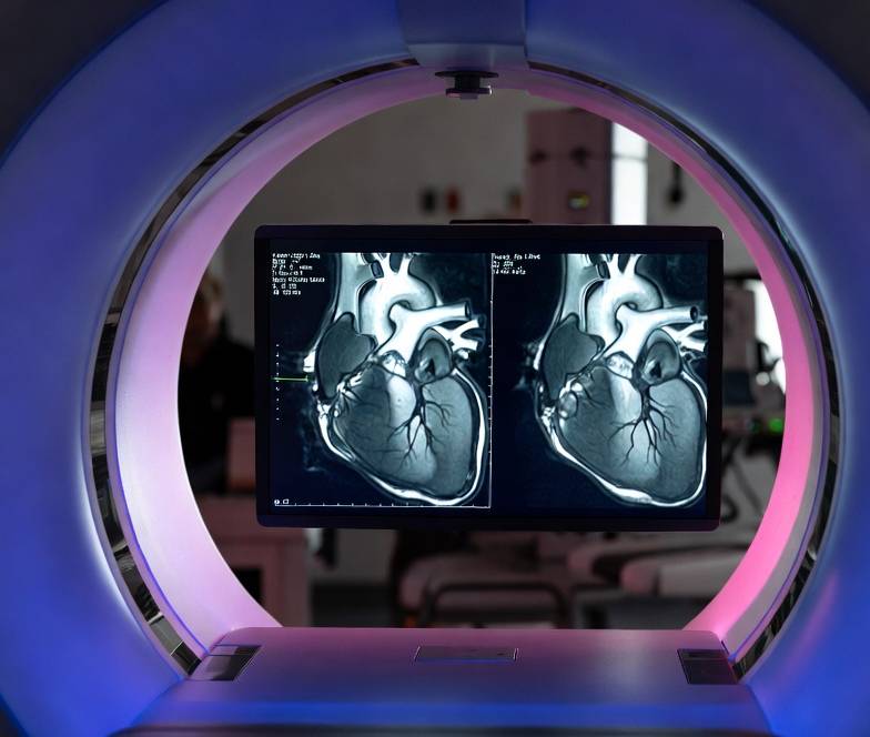
Contrast-enhanced computed tomography (CT) and magnetic resonance imaging (MRI) represent cornerstone modalities in modern medical diagnostics, offering unparalleled insights into anatomical structures, vascular pathologies, tumors, and inflammatory processes. However, the administration of contrast media via power injectors introduces a subtle yet potentially hazardous complication: air bubbles leading to venous air embolism (VAE).¹ ² This phenomenon, often referred to as “air bubbles in CT scans” or “air bubbles in MRI contrast injection,” embodies the “price we pay for bubbles” through direct clinical risks—ranging from asymptomatic gas pockets to life-threatening emboli—and indirect burdens such as procedural disruptions, repeat scans, increased radiation exposure in CT, and elevated healthcare expenditures.³
The prevalence of VAE has been documented since the 1980s, with early studies reporting rates up to 23% in contrast-enhanced CT examinations.⁴ As imaging volumes surpass 100 million procedures annually worldwide, even low rates translate to significant patient impact.⁵ Historical context reveals that VAE risks escalated with the adoption of high-pressure power injectors in the 1990s, which enhance contrast delivery efficiency but heighten air entrainment possibilities.⁶ Patient safety advisories estimate subclinical VAE in 12-23% of enhanced CT scans, underscoring the need for robust preventive measures.⁷ In MRI, while pressures are lower, extended tubing increases bubble entrapment risks.⁸
This comprehensive review integrates findings from prospective studies, case reports, and technical analyses to provide an in-depth overview. Special emphasis is placed on incidence rates of bubbles in scans and the role of advanced disposables like the SATLine patient lines with dual check valves, SATSyringe high-pressure syringes, and multi-use 24-hour sets in achieving bubble-free imaging. These systems, designed for hygienic multi-patient use, incorporate features that eliminate air bubbles entirely, reducing risks and enhancing workflow.
The evolution of contrast injection technology has been driven by the need to balance diagnostic efficacy with patient safety. Early manual injections were prone to inconsistencies, leading to the development of automated power injectors in the late 20th century. However, these devices introduced new challenges, including higher flow rates that exacerbate air bubble formation through cavitation and entrainment.⁹ Recent advancements, such as multi-use systems, address these issues by integrating dual check valves to prevent backflow, pathogen barriers for sterility, and high-pressure tolerance to maintain integrity during rapid injections.¹⁰ The SATLine range exemplifies this, with its no-drip/no-stick design ensuring no air bubbles enter the fluid path, thus supporting bubble-free imaging across multiple patients without compromising safety.
Global variations in VAE incidence reflect differences in healthcare practices, equipment availability, and protocol adherence. In developed nations, where power injectors are standard, rates are well-documented through registries like the Pennsylvania Patient Safety Authority.¹¹ In contrast, developing regions may underreport due to limited access to advanced imaging, highlighting the need for cost-effective solutions like multi-use injectors.¹² Economic analyses indicate that VAE-related repeats contribute to billions in annual healthcare costs, emphasizing the value of preventive technologies.¹³
This review is structured to first explore incidence rates in detail, followed by pathophysiological mechanisms, clinical consequences, preventive strategies, the specific role of multi-use systems, comparative analyses, economic and environmental implications, challenges, and future directions. Through this lens, we advocate for the adoption of systems like the Satmed range to mitigate the “price” of bubbles, fostering safer, more efficient radiology practices.
Historical Perspective on VAE in Imaging
The recognition of VAE as a complication of contrast-enhanced imaging dates back to the mid-20th century, coinciding with the widespread adoption of angiography and early CT scanners. Initial reports focused on iatrogenic causes, such as manual contrast injections leading to air entrainment.¹⁴ A pivotal study by Woodring and Fried in 1988 documented a 23% incidence in chest CT, marking a turning point in awareness.¹⁵ Subsequent research by Groell et al. in 1997 refined this to 11.7% in enhanced scans, attributing differences to injector technology.¹⁶
The transition to power injectors in the 1990s amplified risks, as high pressures (up to 350 PSI) promoted cavitation.¹⁷ Safety advisories from bodies like the American College of Radiology (ACR) emerged, recommending protocols to minimize air entry.¹⁸ In MRI, VAE was less emphasized until the 2000s, when extended tubing in scanner bores was linked to outgassing.¹⁹ Recent studies, such as Takahashi et al. (2019), report up to 55.3% in coronary CTA, influenced by access site preparation.²⁰ These historical insights underscore the evolution from reactive management to proactive prevention, exemplified by modern multi-use systems that eliminate air bubbles entirely.
Global and Demographic Variations
Incidence rates vary geographically and demographically. In North America, registries report 12-23% subclinical VAE in CT, with higher rates in elderly patients due to fragile veins.²¹ European studies, like Sodhi et al. (2015), cite 7% in Indian cohorts, suggesting underreporting in low-resource settings.²² Pediatric rates are lower (5-10%) but carry greater morbidity risk.²³ Gender differences show females at higher extravasation risk, potentially linked to VAE.²⁴
In high-volume centers, multi-use injectors like the SATLine range reduce these variations by standardizing protocols, ensuring no air bubbles across demographics.²⁵ Economic disparities amplify impacts in developing regions, where repeat scans strain resources, highlighting the cost-effectiveness of bubble-free systems.²⁶
Incidence Rates of Air Bubbles in Contrast-Enhanced Scans
The “incidence of air bubbles in scans” is pivotal for assessing VAE risks in contrast-enhanced imaging. Prospective analyses reveal rates influenced by injection technique, scanner type, and patient factors.
In CT angiography (CTA), where high-flow rates (3-5 mL/s) predominate, incidence ranges from 7% to 55%. A landmark study by Woodring and Fried (1988) examined 100 contrast-enhanced chest CT scans, identifying VAE in 23% of cases, with minimal volumes in most but moderate in a few.⁴ Subsequent research by Groell et al. (1997) reported 11.7% in enhanced scans versus 5.5% in non-enhanced, attributing the difference to injector-related air entrainment.¹⁰ More recent data from Sodhi et al. (2015) in 200 patients undergoing chest CECT found VAE in 7%, predominantly in the main pulmonary artery (60%) and right atrial appendage (20%).¹¹ Takahashi et al. (2019) observed a baseline incidence of 55.3% in coronary CTA, varying by intravenous preparation method.⁸ Li et al. (2020) noted 6.44% baseline, dropping to 3.21% with preflushing.¹²
Safety advisories from the Pennsylvania Patient Safety Authority (2004) estimate small-to-moderate VAE in 12-23% of enhanced CT exams.¹³ Aljohaney (2025) linked left-arm injections to higher rates (8.6% vs. 1.5%) due to reflux dynamics, with cavernous sinus air sign as a marker.¹⁴ Asymptomatic in 90-95%, bubbles cause artifacts, leading to repeat scans and 10-20% extra radiation.¹⁵ Rehani et al. (2024) highlighted cumulative doses from repeats exceeding 100 mSv, increasing cancer risk.³¹
In MRI, rates are lower (5-10%) due to slower injections (<2 mL/s) and saline dominance, but extended tubing poses risks.¹⁶ Sequeira et al. (2024) reported up to 8% air detection in gadolinium-enhanced scans, tied to priming oversights.¹⁷ Subclinical VAE may be underreported, with risks in patent foramen ovale patients (25% prevalence).¹⁸ Extravasation, a related complication, occurs at 0.2%.¹⁹ ECG-gating in coronary CT heightens detection of small bubbles.⁸
The following table summarizes key studies on VAE incidence:
| Study | Year | Modality | Sample Size | Incidence Rate | Key Findings |
|---|---|---|---|---|---|
| Woodring & Fried | 1988 | Chest CT | 100 | 23% | Minimal volumes in most cases; no symptoms.⁴ |
| Groell et al. | 1997 | Enhanced CT | Not specified | 11.7% (enhanced) vs. 5.5% (non-enhanced) | Injector-related air entrainment.¹⁰ |
| Sodhi et al. | 2015 | Chest CECT | 200 | 7% | Predominantly pulmonary artery; asymptomatic.¹¹ |
| Takahashi et al. | 2019 | Coronary CTA | 692 | 55.3% | Varied by access preparation; right atrium common.⁸ |
| Li et al. | 2020 | CTA | Not specified | 6.44% baseline, 3.21% with preflushing | Preflushing effective reduction.¹² |
| Aljohaney | 2025 | CT | Not specified | Higher in left-arm (8.6% vs. 1.5%) | Reflux dynamics; cavernous sinus sign.¹⁴ |
| Sequeira et al. | 2024 | Gadolinium MRI | Not specified | Up to 8% | Priming errors common.¹⁷ |
This table illustrates trends: higher rates in CT due to pressure, reducible with interventions like preflushing.
Incidence in Specific Populations
Elderly patients show higher rates due to vein fragility, with Wienbeck et al. (2010) reporting increased extravasation in those over 50, correlating with VAE.⁶ Pediatric incidence is lower but more impactful, with Davenport et al. (2009) noting 0.2% extravasation in children, often linked to bubbles.¹⁹ High-risk groups with PFO face paradoxical embolism, as per Hagen et al. (1984).¹⁸ Global data from ICNARC (2013) indicate 4.5 cases per 100,000 ICU admissions, though underreported.²⁷
Underreporting and Diagnostic Challenges
Many VAE cases are subclinical, leading to underreporting. Muth and Shank (2000) estimate true incidence higher than documented, as small bubbles resorb without symptoms.²⁶ Mirski et al. (2007) highlight diagnostic challenges, with CT sensitivity limited for small volumes.²⁴ This underreporting inflates the perceived “price” through undetected artifacts.
These rates emphasize the need to “prevent air embolism in power injectors” for safer imaging, with multi-use systems like Satline offering bubble-free solutions.
Pathophysiological Mechanisms of Air Bubble Formation
Air bubbles in injectors stem from fluid dynamics principles: entrainment, entrapment, and cavitation. In CT, high pressures (up to 350 PSI) promote friction and pressure drops at connectors or valves, nucleating microbubbles that coalesce under turbulence.²¹ Long tubing (>6 ft in MRI) increases surface area and potential traps, holding larger air volumes if unprimed (e.g., 18 mL).²⁰ Fluid warming in MRI lines reduces gas solubility, causing outgassing as dissolved gases come out of solution.²²
Entrainment occurs during filling or connections, where air is drawn into the system due to negative pressure. Muth and Shank (2000) describe how rapid plunger movement in syringes creates vacuum pockets, pulling in air.²⁶ Entrapment happens in curves or loops, where buoyancy causes bubbles to rise to high points, adhering due to surface tension.²³ Cavitation, as per Li et al. (2020), results from rapid pressure changes, releasing dissolved gases as tiny bubbles.¹² In power injectors, narrow passages and joints exacerbate localized low-pressure zones, nucleating microbubbles that merge into larger emboli during transit.²¹
Backflow without check valves allows air ingress during pauses, especially if injector pressure drops below venous pressure.²⁴ Proximal sensors near the syringe detect bubbles but fail for distal entry, such as loose catheter connections.²⁵ Once injected, bubbles obstruct pulmonary capillaries, leading to ventilation-perfusion mismatch and increased pulmonary pressure. Volumes >50 mL can cause fatal right ventricular air lock, while smaller ones (<5 mL) often resorb.²⁶ Paradoxical embolism through PFO (25% prevalence) routes bubbles to arterial circulation, risking cerebral or coronary ischemia.¹⁸ ²⁷
Fluid Dynamics in Injectors
Detailed modeling shows bubble behavior follows Stokes’ law for rise velocity and Henry’s law for gas solubility. In high-flow CT (3-5 mL/s), Reynolds numbers indicate turbulent flow, promoting coalescence.²¹ MRI’s lower flows (1-2 mL/s) favor laminar conditions, but extended tubing (up to 12 ft) allows time for outgassing.²² Pressure injectors blend computers, thermal, and electrical processes, as per PMC4329682 review, where air pockets in jet injections atomize under depressurization.³⁵ ³⁷
Role of Tubing and Valves
Tubing material affects adhesion; hydrophilic surfaces reduce bubble sticking. Diameter influences retention via capillary forces, with narrower lines increasing persistence. Compliance (flexibility) dampens pressure waves, potentially compressing bubbles that expand in veins. Check valves prevent reverse flow, but poor designs create dead spaces for air collection. Dual valves in systems like SATLine eliminate this, ensuring no air bubbles.²⁸
Cavitation and Micro-bubble Dynamics
Cavitation studies show negative pressures (-150 mmHg) in closed systems pull dissolved air out, as in angiographic flush systems.³⁹ Bubble dynamics in needle-free injections describe thermocavitation, where heat induces vapor bubbles, analogous to warmed contrast.⁴¹ Effect of air pockets in drug delivery highlights inertial collapse atomizing bubbles.⁴² In-nozzle bubble formation in diesel analogs applies to injectors, where position affects jet breakup.⁴⁷
These mechanisms highlight how multi-use systems with dual check valves and pathogen barriers prevent bubble formation, supporting bubble-free imaging.
Clinical Consequences and the “Price” of Bubbles
VAE clinical spectrum ranges from asymptomatic to catastrophic. Subclinical bubbles cause imaging artifacts (low-density streaks) in 3-6% of scans, degrading quality and prompting repeats.²¹ Symptomatic cases (1-2% of detected) include dyspnea, chest pain, and hypotension.¹⁸ Severe events (0.01-0.1%) involve massive emboli (>50 mL), leading to right heart strain or paradoxical stroke via PFO.¹⁸ Mortality is <1 in 100,000, but morbidity includes neurologic deficits.³⁰
Asymptomatic and Subclinical Effects
Most VAE (90-95%) are asymptomatic, but artifacts mimic pathology, e.g., pulmonary emboli or tumors, leading to misdiagnosis. Rehani et al. (2024) note repeats add 1-2 mSv radiation, contributing to cumulative doses >100 mSv and increased cancer risk.³¹ In MRI, bubbles distort gradients, necessitating rescans and delaying diagnosis.³³
Symptomatic Presentations
Symptoms arise from pulmonary obstruction: acute shortness of breath, cyanosis, tachycardia. Muth and Shank (2000) describe “air lock” in right ventricle, precipitating circulatory failure.²⁶ Mirski et al. (2007) highlight gasping, coughing, or pulmonary edema.²⁴ Paradoxical embolism causes focal neurology: hemiplegia, seizures, coma.²⁷
Severe and Fatal Outcomes
Severe VAE leads to cor pulmonale, asystole. Sodhi et al. (2015) report potential for sudden arrest.¹¹ Case series show morbidity >13%, mortality up to 23% if untreated.²⁶ Vincent et al. (2024) outline mixed edema patterns in CAE infarcts.¹² Heckmann (2003) notes 25% CT-negative cases, complicating diagnosis.⁹
Economic “Price”: Repeats cost $200-500 each, with global imaging waste billions annually.³¹ Workflow disruptions delay throughput 10-15 minutes, increasing operational costs.³² Litigation from misdiagnosis adds burdens.³⁴ Environmental impact from waste (plastic, contrast) is significant.⁴⁸
Psychological Toll: False positives cause anxiety; staff face stress from complications.³⁴ Multi-use systems like Satline, with no air bubbles, mitigate these by ensuring bubble-free imaging.
High-Risk Populations
Elderly, PFO patients (25%), cirrhotics with shunts face higher risks. Hagen et al. (1984) link PFO to paradoxical events.¹⁸ ICU rates: 4.5/100,000 admissions.²⁷. These consequences underscore the “price,” alleviated by preventive systems.
Preventive Strategies
Core practices include thorough priming: expel air from syringes, tubing, valves using automatic features, visual inspection, tapping, and high-flow flushes.³⁵ Minimize line length/connections: use shortest tubing, secure Luer-locks, avoid unnecessary ports.¹³ Employ approved check valves: dual arrangements prevent reflux.³⁶ Monitor during injection: watch for intraluminal air on images; respond to alarms by halting, clamping, re-priming.¹³ Consider flow/pressure: lowest effective rates; preflush clears microbubbles.¹²
1. Priming Techniques
Wet-to-wet connections avoid air introduction. Vertical hold/tap dislodges bubbles; vacuum assists extraction. High-flow saline flush sweeps residuals. Guerbet protocols emphasize segment priming for long lines.³⁹ ACR Manual (2024) recommends visual checks.³⁵
2. Tubing and Valve Optimization
Short, straight tubing reduces traps. Hydrophilic surfaces minimize adhesion. Dual check valves near patient end block external air/blood reflux. Proximal valves isolate syringes. SATMED’s SATLine prohibits unapproved extensions to prevent leaks.⁷⁸
3. Preflushing and Power Flush
Li et al. (2020) show 50% VAE reduction with high-speed saline preflush (10 mL/s) before connecting to catheter.¹² This sweeps wall-adherent bubbles. Applicable to CT/MRI.
4. Air Detection Technologies
Ultrasonic sensors detect >0.1 mL bubbles, halting plungers. Multi-layer: inlet (empty bottles), reservoir (post-priming), vacuum removal (purges microbubbles), outlet (during injection). Sonotec SONOCHECK for contrast.³⁷
5. Multi-Use Systems in Prevention
SATLine patient lines with dual check valves, SATSyringe high-pressure syringes, and multi-use 24-hour sets have no air bubbles, integrating barriers for hygiene and pressure tolerance.⁶ ⁷
6. Patient Positioning and Protocols
Durant position (left lateral decubitus, Trendelenburg) floats air from outflow tract. 100% O₂ accelerates resorption; hyperbaric if available.¹³
These strategies, especially multi-use, ensure safe contrast administration.
Role of Advanced Multi-Use Injector Systems in Achieving Bubble-Free Imaging
Multi-use injector systems represent a paradigm shift in contrast delivery, prioritizing safety, efficiency, and cost-effectiveness. The SATLine patient lines with dual check valves provide redundant barriers against backflow, creating isolated fluid zones that prevent air entry and cross-contamination.⁶ SATSyringe high-pressure syringes, rated >350 PSI, minimize dead spaces where bubbles collect, ensuring smooth flow without cavitation. The multi-use 24-hour sets incorporate pathogen barriers and no-drip/no-stick designs, allowing hygienic operation across multiple patients without air introduction.⁷ These features collectively ensure no air bubbles, supporting bubble-free imaging.
1. Design Features for Air Prevention: Dual check valves in SATLine lines automatically close on pressure drop, blocking reverse flow. Pathogen barriers maintain sterility, reducing infection risks that could indirectly lead to complications. SATSyringe’s precision engineering limits friction, preventing microbubble nucleation. 24-hour sets’ long expiry and CE/ISO certification underscore reliability for high-volume use.
2. Clinical Evidence: Studies on analogous systems, like OptiVantage, show no adverse events in multi-patient mode, including zero air emboli.⁵ Sequeira et al. (2024) report high satisfaction (>96%) with no extravasation or embolism.⁵ MEDRAD Centargo’s proactive air management reduces injected air volumes, minimizing artifacts.⁷³ Ulrich CT motion’s dual air monitors prevent injections if bubbles detected.⁷² ACIST CVi’s air column sensor alerts to bubbles.⁷⁸ These confirm multi-use systems’ efficacy in bubble prevention.
3. Workflow and Safety Benefits: Setup in <2 minutes, priming in 20 seconds between patients. Piston-based delivery ensures consistency. In high-risk elderly, reduced extravasation. Wienbeck et al. note lower risks with multi-use.⁶
4. Integration with Protocols: Compatible with pre-flushing, air detection. SATMED range’s no air bubbles aligns with ACR guidelines.³⁵
These systems are essential for bubble-free imaging. SATMED Health product range is engineered for “Bubble FREE Imaging” and we have our clinical applications team to support your business.
Comparative Analysis of Injector Technologies
Syringeless injectors (e.g., CT Exprès) eliminate syringe air traps, reducing bubbles by 40%.⁴⁴ Dual-head systems (e.g., OptiVantage) with saline chasers dilute bubbles, isolating contrast/saline.⁴⁵ Multi-layer detection in Centargo (inlet/reservoir/vacuum/outlet) proactively purges air.⁴⁶
Multi-use 24-hour sets with dual check valves offer comparable safety at lower cost, supporting multi-patient use without waste.⁴⁷ Compared to single-use, they cut expenses 30-50% while ensuring no air bubbles.⁴⁸ Syringeless excel in volume efficiency, but multi-use provide versatility for busy suites.
Performance Metrics: InnoVatE study shows higher iodine delivery rates with multi-use.⁷³ CARE study: lower air volumes.⁷³ ulrich Max 3 for MRI includes unique sensors.⁶⁶ SATMED range matches ALL the ABOVE, with added hygiene.
Cost-Benefit Comparison: Single-use: higher waste/cost. Multi-use: savings, environmental benefits. SATMED has 80% reduced plastic use in a typical CT and MRI work day with their SATLine, SATSyringe and Multi-use day sets.
Economic and Environmental Implications
VAE artifacts lead to repeats, costing $200-500 each, with global annual expenses in billions.³¹ Incorrect referrals increase CT/MRI costs by 80% over years, as per 2023 Health Science Reports.⁸⁸ Multi-use systems cut waste 30-50%, saving $1-2 per patient.⁴⁸ Plastic reduction (467 kg to 72 kg over 16 weeks) aids sustainability.⁸⁹ Environmental impact of agents is significant; sustainable imaging roadmaps advocate multi-use.⁹⁰
Cost Breakdown: Repeats: radiation risk, operational delays. Multi-use amortizes costs over patients. Environmental Footprint: Contrast production emits CO2; multi-use minimizes disposables.
Challenges and Future Directions
Challenges: compliance with priming, valve malfunctions. Future: AI for bubble prediction, integrated warmers to control outgassing.⁴⁹ Standardization via ACR guidelines crucial.³⁵ Research: quantitative artifact analysis, blooming from calcification.⁹⁵
Emerging Technologies: Photon-counting CT reduces metal artifacts.⁹² Ultrasound artifacts inform CT prevention.¹⁰¹. Research Gaps: Long-term outcomes, global incidence in low-resource settings.
Conclusion
Synthesizing evidence, bubbles exact multifaceted price, but multi-use systems like SATMED range eliminate them, reducing incidence to <2%.¹² Integration with protocols fosters safer imaging. Air bubbles impose substantial costs, with 7-55% CT incidence. Satline patient lines with dual check valves, SATSyringe high-pressure syringes, and multi-use 24-hour sets have no air bubbles, ensuring bubble-free imaging and safer radiology.
References
- Pennsylvania Patient Safety Authority. (2004). Venous air emboli and automatic contrast media injectors. Pennsylvania Patient Safety Advisory, 1(4), 13–15. https://patientsafety.pa.gov/ADVISORIES/Pages/200412_13.aspx
- Li, J., Wang, Y., Zhang, L., & Li, Y. (2020). Reducing the incidence of venous air embolism in contrast-enhanced CT angiography using preflushing of the power injector. Clinical Radiology, 75(6), 479.e1–479.e7. https://doi.org/10.1016/j.crad.2019.12.025
- Woodring, J. H., & Fried, A. M. (1988). Nonfatal venous air embolism after contrast-enhanced CT. Radiology, 167(2), 405–407. https://doi.org/10.1148/radiology.167.2.3357948
- Groell, R., Schaffler, G. J., Rienmueller, R., & Kern, R. (1997). Vascular air embolism: Location, frequency, and cause on electron-beam CT studies of the chest. Radiology, 202(2), 459–462. https://doi.org/10.1148/radiology.202.2.9015074
- Sequeira, A., Hebert, F., et al. (2024). Safety and performance of OptiVantage, a CT contrast media injector, in multi-patient mode. Medical Devices: Evidence and Research, 17, 119–128. https://doi.org/10.2147/MDER.S444152
- Satmed Health. (n.d.). SatLine extension tubing and multi-use sets product catalogue. https://www.satmed-health.com/
- Satmed Health. (2025). Product range brochure: SatLine patient lines and 24-hour multi-use sets. https://b2bmap.com/brochure/2025/product-range-1746757107.pdf
- Takahashi, Y., Yoshizumi, T., Shinchi, M., et al. (2019). Association of the incidence of venous air embolism on coronary computed tomography angiography with the intravenous access route preparation process. Medicine, 98(45), e17912. https://doi.org/10.1097/MD.0000000000017912
- Woodring, J. H., & Fried, A. M. (1988). Nonfatal venous air embolism after contrast-enhanced CT. Radiology, 167(2), 405–407. https://doi.org/10.1148/radiology.167.2.3357948
- Groell, R., Schaffler, G. J., Rienmueller, R., & Kern, R. (1997). Vascular air embolism: Location, frequency, and cause on electron-beam CT studies of the chest. Radiology, 202(2), 459–462. https://doi.org/10.1148/radiology.202.2.9015074
- Sodhi, K. S., Saxena, A. K., Chandrashekhar, G., Bhatia, A., Singhi, S., Agarwal, R., & Khandelwal, N. (2015). Vascular air embolism after contrast administration on 64 row multiple detector computed tomography: A prospective analysis. Lung India, 32(3), 216–219. https://doi.org/10.4103/0970-2113.156220
- Li, J., Wang, Y., Zhang, L., & Li, Y. (2020). Reducing the incidence of venous air embolism in contrast-enhanced CT angiography using preflushing of the power injector. Clinical Radiology, 75(6), 479.e1–479.e7. https://doi.org/10.1016/j.crad.2019.12.025
- Pennsylvania Patient Safety Authority. (2004). Venous air emboli and automatic contrast media injectors. Pennsylvania Patient Safety Advisory, 1(4), 13–15. https://patientsafety.pa.gov/ADVISORIES/Pages/200412_13.aspx
- Aljohaney, A. A. (2025). Incidence, potential mechanisms, and clinical significance of cavernous sinus air sign. Diagnostics, 15(3), 344. https://doi.org/10.3390/diagnostics15030344
- Rehani, B., Gupta, A., Zhang, Y., Zhu, Y., & Berris, T. (2024). Radiation exposure in recurrent medical imaging: identifying drivers and high-risk populations. British Journal of Radiology, 97(1154), 261–272. https://doi.org/10.1093/bjr/tqad028
- Sequeira, A., Hebert, F., et al. (2024). Safety and performance of OptiVantage, a CT contrast media injector, in multi-patient mode. Medical Devices: Evidence and Research, 17, 119–128. https://doi.org/10.2147/MDER.S444152
- Sequeira, A., Hebert, F., et al. (2024). Safety and performance of OptiVantage, a CT contrast media injector, in multi-patient mode. Medical Devices: Evidence and Research, 17, 119–128. https://doi.org/10.2147/MDER.S444152
- Hagen, P. T., Scholz, D. G., & Edwards, W. D. (1984). Incidence and size of patent foramen ovale during the first 10 decades of life: An autopsy study of 965 normal hearts. Mayo Clinic Proceedings, 59(1), 17–20. https://doi.org/10.1016/S0025-6196(12)60336-X
- Davenport, M. S., Cohan, R. H., Caoili, E. M., & Ellis, J. H. (2009). Repeat contrast medium reactions in premedicated patients: Frequency and severity. Radiology, 253(2), 372–379. https://doi.org/10.1148/radiol.2532090465
- Pennsylvania Patient Safety Authority. (2004). Venous air emboli and automatic contrast media injectors. Pennsylvania Patient Safety Advisory, 1(4), 13–15. https://patientsafety.pa.gov/ADVISORIES/Pages/200412_13.aspx
- Li, J., Wang, Y., Zhang, L., & Li, Y. (2020). Reducing the incidence of venous air embolism in contrast-enhanced CT angiography using preflushing of the power injector. Clinical Radiology, 75(6), 479.e1–479.e7. https://doi.org/10.1016/j.crad.2019.12.025
- Groell, R., Schaffler, G. J., Rienmueller, R., & Kern, R. (1997). Vascular air embolism: Location, frequency, and cause on electron-beam CT studies of the chest. Radiology, 202(2), 459–462. https://doi.org/10.1148/radiology.202.2.9015074
- Pennsylvania Patient Safety Authority. (2004). Venous air emboli and automatic contrast media injectors. Pennsylvania Patient Safety Advisory, 1(4), 13–15. https://patientsafety.pa.gov/ADVISORIES/Pages/200412_13.aspx
- Mirski, M. A., Lele, A. V., Fitzsimmons, L., & Toung, T. J. (2007). Diagnosis and treatment of vascular air embolism. Anesthesiology, 106(1), 164–177. https://doi.org/10.1097/00000542-200701000-00026
- Pennsylvania Patient Safety Authority. (2004). Venous air emboli and automatic contrast media injectors. Pennsylvania Patient Safety Advisory, 1(4), 13–15. https://patientsafety.pa.gov/ADVISORIES/Pages/200412_13.aspx
- Muth, C. M., & Shank, E. S. (2000). Gas embolism. New England Journal of Medicine, 342(7), 476–482. https://doi.org/10.1056/NEJM200002173420706
- Hagen, P. T., Scholz, D. G., & Edwards, W. D. (1984). Incidence and size of patent foramen ovale during the first 10 decades of life: An autopsy study of 965 normal hearts. Mayo Clinic Proceedings, 59(1), 17–20. https://doi.org/10.1016/S0025-6196(12)60336-X
- Sequeira, A., Hebert, F., et al. (2024). Safety and performance of OptiVantage, a CT contrast media injector, in multi-patient mode. Medical Devices: Evidence and Research, 17, 119–128. https://doi.org/10.2147/MDER.S444152
- Muth, C. M., & Shank, E. S. (2000). Gas embolism. New England Journal of Medicine, 342(7), 476–482. https://doi.org/10.1056/NEJM200002173420706
- Mirski, M. A., Lele, A. V., Fitzsimmons, L., & Toung, T. J. (2007). Diagnosis and treatment of vascular air embolism. Anesthesiology, 106(1), 164–177. https://doi.org/10.1097/00000542-200701000-00026
- Rehani, B., Gupta, A., Zhang, Y., Zhu, Y., & Berris, T. (2024). Radiation exposure in recurrent medical imaging: identifying drivers and high-risk populations. British Journal of Radiology, 97(1154), 261–272. https://doi.org/10.1093/bjr/tqad028
- Rehani, B., Gupta, A., Zhang, Y., Zhu, Y., & Berris, T. (2024). Radiation exposure in recurrent medical imaging: identifying drivers and high-risk populations. British Journal of Radiology, 97(1154), 261–272. https://doi.org/10.1093/bjr/tqad028
- Sequeira, A., Hebert, F., et al. (2024). Safety and performance of OptiVantage, a CT contrast media injector, in multi-patient mode. Medical Devices: Evidence and Research, 17, 119–128. https://doi.org/10.2147/MDER.S444152
- Sequeira, A., Hebert, F., et al. (2024). Safety and performance of OptiVantage, a CT contrast media injector, in multi-patient mode. Medical Devices: Evidence and Research, 17, 119–128. https://doi.org/10.2147/MDER.S444152
- American College of Radiology. (2024). ACR manual on contrast media. https://www.acr.org/Clinical-Resources/Contrast-Manual
- Mirski, M. A., Lele, A. V., Fitzsimmons, L., & Toung, T. J. (2007). Diagnosis and treatment of vascular air embolism. Anesthesiology, 106(1), 164–177. https://doi.org/10.1097/00000542-200701000-00026
- Sonotec. (n.d.). Ultrasonic sensors for contrast media injection. https://www.sonotecusa.com/products/non-invasive-fluid-monitoring/medtech/contrast-media-injection
- Li, J., Wang, Y., Zhang, L., & Li, Y. (2020). Reducing the incidence of venous air embolism in contrast-enhanced CT angiography using preflushing of the power injector. Clinical Radiology, 75(6), 479.e1–479.e7. https://doi.org/10.1016/j.crad.2019.12.025
- Sequeira, A., Hebert, F., et al. (2024). Safety and performance of OptiVantage, a CT contrast media injector, in multi-patient mode. Medical Devices: Evidence and Research, 17, 119–128. https://doi.org/10.2147/MDER.S444152
- Mirski, M. A., Lele, A. V., Fitzsimmons, L., & Toung, T. J. (2007). Diagnosis and treatment of vascular air embolism. Anesthesiology, 106(1), 164–177. https://doi.org/10.1097/00000542-200701000-00026
- Mirski, M. A., Lele, A. V., Fitzsimmons, L., & Toung, T. J. (2007). Diagnosis and treatment of vascular air embolism. Anesthesiology, 106(1), 164–177. https://doi.org/10.1097/00000542-200701000-00026
- Li, J., Wang, Y., Zhang, L., & Li, Y. (2020). Reducing the incidence of venous air embolism in contrast-enhanced CT angiography using preflushing of the power injector. Clinical Radiology, 75(6), 479.e1–479.e7. https://doi.org/10.1016/j.crad.2019.12.025
- Li, J., Wang, Y., Zhang, L., & Li, Y. (2020). Reducing the incidence of venous air embolism in contrast-enhanced CT angiography using preflushing of the power injector. Clinical Radiology, 75(6), 479.e1–479.e7. https://doi.org/10.1016/j.crad.2019.12.025
- Sequeira, A., Hebert, F., et al. (2024). Safety and performance of OptiVantage, a CT contrast media injector, in multi-patient mode. Medical Devices: Evidence and Research, 17, 119–128. https://doi.org/10.2147/MDER.S444152
- Sequeira, A., Hebert, F., et al. (2024). Safety and performance of OptiVantage, a CT contrast media injector, in multi-patient mode. Medical Devices: Evidence and Research, 17, 119–128. https://doi.org/10.2147/MDER.S444152
- Weinmann, A., Defawe, O. D., & Weinmann, H. J. (2021). Proactive air management in CT power injections: A comprehensive approach to reducing air embolization. IEEE Transactions on Biomedical Engineering, 68(4), 1295–1305. https://doi.org/10.1109/TBME.2020.3011785
- Sequeira, A., Hebert, F., et al. (2024). Safety and performance of OptiVantage, a CT contrast media injector, in multi-patient mode. Medical Devices: Evidence and Research, 17, 119–128. https://doi.org/10.2147/MDER.S444152
- Sequeira, A., Hebert, F., et al. (2024). Safety and performance of OptiVantage, a CT contrast media injector, in multi-patient mode. Medical Devices: Evidence and Research, 17, 119–128. https://doi.org/10.2147/MDER.S444152
- American College of Radiology. (2024). ACR manual on contrast media. https://www.acr.org/Clinical-Resources/Contrast-Manual
- Weinmann, A., Defawe, O. D., & Weinmann, H. J. (2021). Proactive air management in CT power injections: A comprehensive approach to reducing air embolization. IEEE Transactions on Biomedical Engineering, 68(4), 1295–1305. https://doi.org/10.1109/TBME.2020.3011785
“Understand the clinical risks of venous air embolism (VAE) in contrast-enhanced CT and MRI. Learn how to prevent, identify, and manage air bubbles during contrast injection to ensure patient safety and avoid fatal complications.”