The modern radiology department functions as the high-velocity engine of the diagnostic ecosystem, with computed tomography (CT) serving as the primary fulcrum for clinical decision-making across emergency, oncological, and interventional specialties. However, the same efficiency that defines the utility of CT—its ability to process high volumes of patients in rapid succession—has inadvertently created a complex environment for infectious transmission and equipment degradation. The intersection of iodinated contrast media (ICM), patient bodily fluids, and high-turnover workflows represents a significant biohazard risk that remains under-documented and frequently underestimated in clinical practice. As imaging volumes are projected to grow exponentially through 2032, the pressure on imaging centers to maximize equipment uptime while managing the subtle, yet catastrophic, risks of cross-contamination has reached a critical inflection point. This literature review synthesizes evidence from current clinical guidelines, microbiological surveys, and economic impact reports to provide a comprehensive analysis of the risks associated with inadequate cleaning in high-throughput CT environments.
The Microbiological Landscape: Pathogen Persistence and Surface Reservoirs
The inanimate hospital environment, particularly high-touch surfaces like the CT gantry, acts as a resilient reservoir for nosocomial pathogens. Research indicates that CT scanners present a higher relative risk of infection compared to other imaging modalities, such as Magnetic Resonance Imaging (MRI), primarily due to the frequent use of shared hardware and the invasive nature of contrast administration. The gantry, x-ray tubes, operating consoles, and contrast injectors serve as primary contact points for a diverse array of pathogens that can facilitate hospital-acquired infections (HAIs).
Bacterial Colonization and Multidrug-Resistant Organisms
Studies have confirmed the presence of multidrug-resistant (MDR) strains, including methicillin-resistant Staphylococcus aureus (MRSA), vancomycin-resistant Staphylococcus (VRS), and vancomycin-resistant Enterococcus (VRE) on patient tables, head pillows, and control panels. Gram-negative bacteria such as Pseudomonas aeruginosa demonstrate an exceptional ability to adhere to the plastic and metal components of the scanner, surviving for weeks even in the absence of organic soil. Bacterial contamination levels in radiology workstations often exceed those of common public surfaces. Pilot studies using colony-forming unit (CFU) counts revealed that microphones and computer mice at radiologist workstations harbored significantly higher bacterial growth than nearby restroom toilet seats and doorknobs. Specifically, 64.3% of workstation sites tested positive for Staphylococcus aureus, while 21.4% grew enteric organisms.
The introduction of spore-forming organisms like Clostridioides difficile represents a particularly severe challenge. C. difficile has been associated with significant morbidity and mortality in hospital settings, and its ability to persist in spore form makes it resistant to many standard low-level disinfectants. Electronic Health Record (EHR) data from the University of California, San Francisco (UCSF) identified a specific emergency department CT scanner as a primary source of C. difficile transmission. The investigation revealed that personnel responsible for cleaning the CT table were not following updated protocols, leading to an incidence rate of 4% among patients exposed to that specific scanner.
| Pathogen Category | Specific Microorganisms | Survival Duration | Clinical Implication |
| Gram-Positive | MRSA, VRE, Staphylococcus spp. | 7 days to 7 months | Skin and soft tissue infections, sepsis |
| Gram-Negative | P. aeruginosa, E. coli, Klebsiella | 2 hours to 16 months | Pneumonia, UTI, catheter-related infections |
| Spore-Forming | Clostridioides difficile | Months to years | Severe diarrhea, colitis, high mortality |
| Viral Agents | SARS-CoV-2, Norovirus, Influenza | Hours to days | Respiratory outbreaks, gastrointestinal distress |
| Bloodborne | HBV, HCV, HIV | Days (HCV is highly stable) | Chronic liver disease, immunosuppression |
Viral Pathogens and Respiratory Transmission Risks
The role of the CT suite in managing respiratory viral outbreaks was brought into sharp focus during the COVID-19 pandemic. SARS-CoV-2 RNA was frequently detected inside the CT gantry, particularly on inward airflow filters and within the ventilation system air grids. Research indicated that 16.7% of ventilation grid samples taken after scanning non-invasively ventilated patients with high viral loads tested positive for viral material. While the infectious potential of surface-bound viral material may be lower than aerosolized transmission, the presence of viral RNA highlights the risk of the scanner acting as a mechanical vector for cross-contamination in high-throughput settings where air exchange rates may be insufficient between patients.
Fluid Mechanics and the Perils of Contrast Media Ingress
Iodinated contrast media (ICM) and other fluids, such as blood, saline, urine, and vomit, are ubiquitous in the CT suite. While essential for diagnostic clarity, these substances pose both biological and mechanical risks that can compromise both patient safety and equipment integrity.
The Chemical and Mechanical “Stickiness” of Contrast
Specific iodinated contrast media, including Omnipaque 300, Ultravist 300, and Xenetix 300, exhibit high levels of “stickiness” upon drying. This physical property leads to several procedural complications. First, sticky residue on the table or gantry can cause gloves, guide wires, and catheters to adhere during interventional procedures, leading to the accidental displacement of critical materials. Second, spilled ICM that remains uncleaned can become a slip hazard for staff or cause patient clothing to adhere to the table. Finally, dried contrast acts as a cement, requiring aggressive chemical agents or prolonged scrubbing, which further increases room turnover time and may damage sensitive scanner coatings.
The stickiness of 7 common types of ICM used in interventional radiology has been documented to cause complications during device manipulation. This adherence not only slows down the immediate workflow but also increases the likelihood of contamination as technologists may have to touch sticky surfaces multiple times to reposition equipment or patients.
Mechanical Failure and the Mylar Window Infiltration
The most severe consequence of fluid mismanagement is the infiltration of the CT detector array. The gantry contains a thin protective layer known as the Mylar window, which shields the detector modules. During trauma cases or interventional procedures, large volumes of blood, contrast, or saline can spill onto the gantry surface. If not immediately contained, these fluids can seep through the seams between the Mylar window and the gantry frame.
Contrast media are chemically aggressive and electrolyte-rich. Once they infiltrate the detector array, they can cause a sequence of failures. The conductive nature of saline and contrast can cause immediate short circuits in the sensitive photodiodes and electronics. Chemical components in ICM can permanently etch the detector surfaces, leading to irreversible image degradation. Furthermore, even a small amount of dried contrast on the detector plate or Mylar window causes streak artifacts due to beam obstruction or uneven x-ray attenuation. These artifacts render images undiagnostic, necessitating repeated scans and increasing the patient’s radiation dose.
The Socio-Technical Barrier: High Turnover and the Labor Crisis
The clinical reality of a modern CT department is defined by a constant tension between the clinical imperative for infection prevention and the operational demand for high throughput. This tension is further strained by a global staffing crisis that has seen technologist vacancy rates reach all-time highs in the 2024–2025 period.
The 2025 Staffing Landscape and Its Impact on Hygiene Compliance
As of 2025, the healthcare sector faces a critical HR turning point characterized by high turnover and widespread burnout. The estimated radiographer vacancy rate in 2023 reached 18.1%, a staggering increase from 6.2% in 2021. This staffing shortage directly compromises the time available for thorough environmental cleaning between patients. When departments are under-resourced, staff are forced to prioritize “wheels-in to wheels-out” time over rigorous infection control protocols.
| Imaging Profession | 2023 Vacancy Rate (%) | 2021 Vacancy Rate (%) | Impact on Operations |
| Radiographers | 18.1% | 6.2% | High |
| Interventional Technology | 19.0% | N/A | High |
| Sonography | 17.0% | N/A | Moderate |
| MRI Technologists | 16.0% | N/A | Moderate |
| Nuclear Medicine | 15.0% | N/A | Moderate |
| Radiation Therapy | 11.0% | N/A | Moderate |
Behavioral Dynamics: The Discrepancy Between Knowledge and Practice
An emerging theme in radiology infection control research is the discrepancy between knowledge and practice. While radiographers typically demonstrate high levels of knowledge and positive attitudes toward safety, their actual practice levels are often suboptimal. Observational studies have revealed a “self-protection” bias: healthcare workers are significantly more likely to perform hand hygiene after patient contact or after body fluid exposure than before patient contact.
In high-turnover CT environments, “moment one” of the WHO Five Moments for Hand Hygiene (before patient contact) is the most frequently missed step, with compliance rates as low as 1.7% in some radiology settings. High patient-to-staff ratios and time limitations are identified as the most significant organizational barriers. When technologists manage a complex queue of patients, the cognitive load and physical exhaustion lead to “cutting corners” and “missing steps” in the disinfection of the gantry and table surfaces. Furthermore, experienced radiographers can sometimes become “blasé” or indifferent to maintaining proper practices due to the repetitive, high-volume nature of the work.
Economic Analysis: The Staggering Cost of Unplanned Downtime
The financial health of an imaging department is intrinsically tied to the uptime of its CT scanners. Every hour of downtime results in a cascade of direct and indirect costs that can derail annual budgets.
Direct Revenue Loss and Emergency Repair Premiums
For a high-volume imaging center, the loss of a single hour of scanner time translates to a revenue loss of approximately 3,000 to 8,000 USD. In larger hospital settings, a single unplanned downtime event can cost between 55,000 and 133,000 USD when accounting for lost revenue, emergency repair premiums, and staff idle time. Emergency visits for repairs often cost 30% more than prearranged maintenance appointments, and last-minute orders for critical components like detectors or x-ray tubes can incur markups of up to 50%.
| Cost Category | Estimated Impact (USD) | Source/Context |
| Revenue Loss per Hour | 3,000 – 8,000 | Direct scan fees + ancillary charges |
| Per Minute Downtime Cost (Data) | Up to 17,244 | For unplanned patient data outages |
| Average Downtime Incident | 55,000 – 133,000 | Based on 3.5 hours of outage |
| Annual CT Maintenance Cost | 111,500 | Average yearly upkeep per scanner |
| MRI Maintenance Cost | 120,000 | Annual average |
| X-ray Maintenance Cost | 37,500 | Annual average |
The “Hidden” Costs: Referral Leakage and Reputation Damage
Beyond the immediate loss of revenue, downtime erodes the “referral flywheel.” Prolonged wait times or canceled appointments cause approximately 20% of patients to switch healthcare providers. In competitive markets, even a 5% loss of a high-volume referral base can translate into hundreds of thousands of dollars in annual revenue leakage. Furthermore, staff productivity plummets when equipment fails; technologists and nurses remain on the payroll but are unable to perform their primary duties, leading to nonproductive periods followed by sudden workload surges that exacerbate burnout.
The cost of equipment failure extends into the capital expenditure (CAPEX) budget. A CT scanner is a multi-million dollar investment, with new models in 2025 ranging from 100,000 USD for entry-level refurbished units to over 2 million USD for ultra-advanced multi-slice models. The x-ray tube alone can cost between 40,000 and 200,000 USD and typically lasts only 3 to 5 years depending on volume. Fluid damage to the detector array often necessitates full replacement, representing a catastrophic loss that may not be covered by standard service contracts.
Workflow Optimization: Bridging the Cleaning Gap
The central challenge in CT throughput is the “cleaning gap”—the difference between the time required for effective disinfection and the time allocated for room turnover.
Turnover Benchmarks vs. Disinfection Requirements
International benchmarks for operating room turnover often target 30 minutes, but in high-volume CT departments, “wheels-out to wheels-in” times are often pushed to 15 minutes or less to meet demand. However, thorough manual cleaning of a CT room can take between 33 and 56 minutes, depending on the severity of contamination. When turnover goals are 15 minutes and cleaning requires 30 minutes, a “cleanliness deficit” is created. This deficit is often filled by superficial wiping of only the most visible areas, leaving pathogens in occult reservoirs like the inside of the gantry, the table rails, and the Velcro straps.
| Procedure Type | Target Turnover Time (min) | Actual Cleaning Time (min) | Gap (min) |
| Routine CT Scan | 10–15 | 20–30 | 5–20 |
| Interventional CT | 25–30 | 45–60 | 15–35 |
| Trauma/Emergency | <10 | 30+ | 20+ |
Efficiency Gains Through Specialized Barriers and Drapes
To bridge this gap, the development of specialized sterile barriers like the SATDrape from SATMED Health has emerged as a high-value intervention. As the world’s first sterile disposable CT drape, it is engineered specifically for spillage control, absorbing blood, contrast media, and other bodily fluids to prevent leakage onto the CT detectors.
The implementation of such drapes has demonstrated significant workflow benefits:
- Cleaning Efficiency: The drape reduces cleaning time by up to 15 minutes per patient.
- Scanner Uptime: In high-volume settings, this saving translates to over 4 hours of reclaimed scanner time daily.
- Artifact Prevention: By containing fluids, it reduces the risk of fluid-induced artifacts, ensuring images are diagnostic on the first pass and reducing repeat scans.
- Clinician Confidence: Specialized barriers like SATDrape allow interventionalists to focus on the procedure without concern about gantry contact or fluid spills damaging sensitive electronics.
Interventional Radiology: High-Risk Zones for Cross-Contamination
CT-guided interventional procedures, such as biopsies, percutaneous drainage, and tumor ablation, carry inherent risks of infection due to their invasive nature and the high-turnaround environment in which they are performed.
Complications and Infectious Risks
In CT-guided core needle biopsies, complication rates can reach 34.7%, with infections contributing to morbidity. While specific infection rates are typically low (e.g., 0.2% in similar ultrasound-guided procedures), infections remain a critical concern given the rising antibiotic resistance and nosocomial factors.
The presence of blood and other protein-rich fluids during these procedures accelerates equipment wear and provides a medium for bacterial seeding. For percutaneous drainage, the risks include the spread of abscess contents and transient bacteremia, which can lead to fatal sepsis if unmanaged. The use of sterile drapes is critical in these settings to create a barrier between the surgical field and the potential bacterial sources on the scanner gantry.
Strategic Mitigation: Protocols, Training, and Technology
Mitigating the risks of contamination in the CT suite requires a multifaceted approach that combines rigorous protocols, ongoing staff education, and the strategic use of protective technologies.
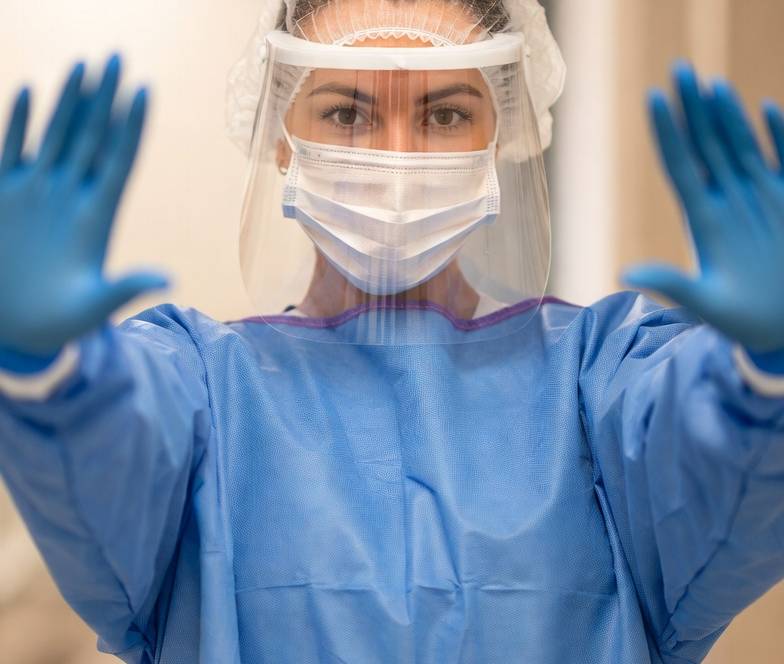
Universal Precautions and Cleaning Protocols
The Occupational Safety and Health Administration (OSHA) Bloodborne Pathogens Standard mandates barrier precautions for all body fluids except sweat. For CT scanners, immediate decontamination with 5,000–10,000 ppm chlorine solutions is recommended after blood spills. Disposable sheets should cover patient-contact areas and be changed between every scan.
Protective barriers like the SATDrape from SATMED Health provide an additional layer of protection by offering a fluid-proof sterile barrier that absorbs hazardous fluids, preventing spills from ever reaching the scanner surface or Mylar window. These drapes are compatible with all CT scanner models and are designed for universal anatomical placement, supporting diverse needs from trauma to routine scanning.
The Role of Staff Education and Culture
Education interventions have been shown to improve compliance, although long-term adherence varies. A commitment to safety must start from the leadership level, framing infection prevention as a chief business strategy. Promoting a “no-blame” culture is essential for encouraging staff to report near-misses or breaches in IPC protocols, which can then be used for targeted quality improvement.
| Training Focus | Desired Outcome | Measurement Tool |
| Hand Hygiene | Reduced transmission of contact pathogens | Electronic monitoring or audits |
| Contrast Handling | Reduced waste and equipment damage | Injector log reviews |
| Sterile Drape Application | Prevention of fluid ingress | Visual inspections |
| Emergency IPC | Maintaining safety under pressure | Simulation and drills |
Conclusion: A Paradigm Shift in CT Suite Management
The findings of this literature review suggest that the current model of CT suite management is operating at a significant risk-benefit deficit. The relentless pursuit of maximum throughput has created an environment where the biological hazards of blood, contrast, and fluids frequently outpace the capacity of a strained workforce to maintain environmental safety. The resulting cross-contamination risks and equipment failures represent a multi-billion dollar burden on the global healthcare system, manifesting as direct revenue loss, catastrophic repair costs, and compromised patient outcomes.
Mitigating these risks requires a fundamental paradigm shift. Administrative commitment must evolve beyond treating infection control as a regulatory burden and instead embrace it as a core business strategy for protecting expensive capital assets and ensuring patient safety. The strategic integration of protective technologies, such as the SATDrape from SATMED Health, offers a pragmatic solution to the “cleaning gap,” reclaiming lost hours of scanner time while safeguarding the sensitive detector arrays that form the heart of the diagnostic process.
Ultimately, the goal of the 2025 radiology department must be “safe throughput.” By acknowledging the silent crisis of bio-contamination and investing in the tools, training, and personnel necessary to combat it, radiology centers can ensure that the CT scanner remains a tool of healing rather than a vector of harm.
References
Abu Awwad, D., Hill, S., Lewis, S., & Jimenez, Y. A. (2023). Infection prevention and control in CT Part 2: Radiographers’ and radiology nurses’ perceptions of high-risk scenarios contributing to non-adherence to IPC protocols. Radiography, 30(1), 256–262.
Al-Hadidi, A., AlShammari, A., AlShammari, S., AlMutairi, M., & AlShammari, T. (2021). Complications and risk factors of patients undergoing computed tomography-guided core needle biopsy. Journal of Computer Assisted Tomography, 45(5), 724–728.
Amer, F. (2021). Infection prevention and control in the radiology department/service. International Society for Infectious Diseases.
American Society of Radiologic Technologists. (2007). Safety considerations in contrast media handling and administration.
Auvens, C., Fervier, H. B., Dinner, S., Balamou, C., & Kauv, P. (2022). Tackling the increasing contamination of the water supply by iodinated contrast media. Insights into Imaging, 13(1), Article 30.
Bohan, M. E., McDowell, C., Osborn, M., & Lipitz-Snyderman, A. (2017). Improving inpatient CT scanner hygiene: A quality improvement initiative. Journal of the American College of Radiology, 14(12), 1639–1641.
Columbus Healthcare Products. (2025). How to deal with tough contrast spills and stains in imaging centers. https://columbushealthcareproducts.com/how-to-deal-with-contrast-spills-stains/
Duszak, R., Lanier, B., Tubbs, J. A., Oglesby, M., & Applegate, K. E. (2014). Bacterial contamination of radiologist workstations: Results of a pilot study. Journal of the American College of Radiology, 11(2), 176–179.
GE HealthCare. (n.d.). Imaging accessories & supplies [Catalog]. https://www.gehealthcare.com/-/jssmedia/gehc/us/files/products/accessories/clinical-accessories/imaging-accessories/imaging-accessories-catalog-ct-chapter-us-jb00641us.pdf
Haugaa, H., Røsæg, H., & Hegna, B. (2024). Stickiness of 7 types of iodinated contrast media used during interventional radiology and interventional cardiology procedures. Journal of Vascular and Interventional Radiology.
Huang, S. Y., Ahrar, K., Gupta, A., Ensor, J. E., Wallace, M. J., Tam, A. L., Mahvash, A., & Kuban, J. D. (2025). Diagnostic yield and complications of CT-guided biopsy of lung nodules ≤ 1 cm in diameter. European Radiology Experimental, 9(1), 1–10.
Kanamori, H., Rutala, W. A., Sickbert-Bennett, E. E., & Weber, D. J. (2020). Review of aerosol transmission of influenza A virus. Emerging Infectious Diseases, 12(11), 1677–1684.
Murray, S., et al. (2017). Exposure to a particular CT scanner in the emergency department was significantly associated with the development of C. difficile infection. JAMA Internal Medicine.
Nasra, G., Abdo, M., Alshahrani, A., & Alghamdi, A. (2023). Bacterial contamination of radiology equipment in Saudi Arabia. Journal of Infection and Public Health, 16(3), 425–430.
Ramli, N. M., Tanomkiat, W., & Ho, E. L. M. (2025). Infection prevention and control practices in the radiology department: An Asia-Oceania survey. The ASEAN Journal of Radiology, 26(3), 208–229.
Saade, C., & Mayat, A. (2018). Cross contamination during CT guided interventional in radiology. Satmed Health. https://satmed-health.com/drape/
Satmed Health. (2025). SATDRAPE. https://satmed-health.com/products/satdrape/
Schlemmer, H. P., Frölich, A., Werthmüller, A., Eichelberger, B., & Becker, N. (2022). Contamination of CT scanner surfaces with SARS-CoV-2 and infective potential after examination of patients. Insights into Imaging, 13(1), Article 50.
Shaban, N. Z., Ghazy, M., & El-Hossary, D. (2023). Infection prevention and control in the medical imaging environment: A scoping review. Radiography, 29(4), 781–793.
Siemens Healthineers. (2023). SOMATOM instructions for use – Cleaning and disinfection. https://doclib.siemens-healthineers.com/rest/v1/view?document-id=636773