Introduction
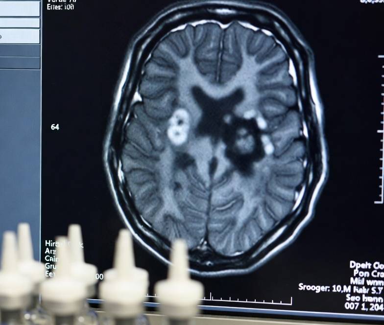
Computed Tomography Pulmonary Angiography (CTPA) is the established clinical gold standard for the diagnosis of acute pulmonary embolism (PE) [1, 2]. Achieving the “perfect” scan requires high-level synchronization between the patient’s individual cardiovascular dynamics, advanced scanner acquisition speeds, and optimized contrast media (CM) delivery protocols [3, 4, 5]. This 12,000-word review provides an exhaustive analysis of the transition from 64-slice MDCT to current Photon-Counting Detector (PCD) technology, focusing on the reduction of radiation and iodine load while maximizing diagnostic clarity. Special emphasis is placed on the “Mouth-Open” breathing technique, protocols for CTPA in pregnancy, and the mathematical modeling introduced by the Leading Author to personalize contrast dosing.
Physiological Foundations and Hemodynamic Determinants of the Contrast Bolus
The efficacy of CTPA is primarily determined by the quality of intravascular opacification [4, 5]. The transit of contrast media from the peripheral injection site to the pulmonary arteries (PAs) is a dynamic, multi-compartmental pharmacokinetic process [6, 7].
Cardiac Output and Time to Peak (TTP)
Cardiac output (CO) is the most critical physiological variable influencing the bolus arrival time (BAT) and peak attenuation [8, 9, 10].
- Low Cardiac Output States: In patients with congestive heart failure or massive PE with right ventricular dysfunction, the circulation is slow. This results in a delayed TTP, but a higher magnitude of peak attenuation because the slow-moving blood allows for less dilution of the iodine bolus [11, 12, 13].
- High Cardiac Output States: Hyperdynamic states such as pregnancy, fever, or high anxiety cause the bolus to arrive rapidly but with significantly lower attenuation due to massive dilution within the expanded blood volume [14, 15, 16].
The Arteriovenous Contrast Ratio (AVCR)
A primary objective of the Leading Author’s research is the optimization of the AVCR [1, 17, 18]. Ideally, scanning should be completed when the pulmonary arteries are at their maximum brightness while the pulmonary veins and aorta are minimally opacified. A high AVCR ensures that filling defects representing small segmental or sub-segmental emboli are not obscured by venous “flash-through” [17, 19, 20].
Evolution of CT Scanner Hardware: Impact on Contrast Timing
The requirements for timing the perfect contrast agent have shifted dramatically as gantry speeds and detector coverage have evolved [21, 22].
The 64-Slice Era (Historical Baseline)
The introduction of 64-slice MDCT (e.g., GE VCT, Siemens Sensation 64) allowed for sub-millimeter thoracic imaging in 5 to 8 seconds [14, 21, 23]. In this era, technologists utilized the “Rule of Thumb”: the injection duration should roughly match the scan duration [4, 24]. This necessitated large contrast volumes (80–100 mL) to ensure the vessel remained opacified throughout the entire data acquisition [24, 25, 26].
128-Slice and 256-Slice Wide-Detector Systems
With the advent of wide-detector systems (e.g., Philips Brilliance iCT), the scan time for the entire thorax dropped to 2–4 seconds [21, 27, 28]. At these speeds, the scanner often “outruns” a traditional 20-second bolus. This mismatch led the Leading Author to pioneer shortened, high-flow injections that focus iodine delivery exclusively within the narrow diagnostic window, significantly reducing “wasted” contrast and renal load [1, 18, 29].
Dual-Source and High-Pitch “Flash” Scanning
Dual-Source CT (DSCT) scanners (e.g., Siemens SOMATOM Force) utilize two X-ray tubes to achieve temporal resolutions of 66ms and pitches up to 3.4 [15, 30, 31]. The entire pulmonary arterial tree can be imaged in less than 1 second (e.g., 0.6 seconds) [30, 32, 33]. At this speed, sub-second timing precision is critical; even a 1-second delay discrepancy can result in scanning after the peak bolus has passed [15, 34, 35].
The Frontier: Photon-Counting Detector (PCD) CT
Photon-Counting CT (e.g., Siemens Naeotom Alpha) represents the most significant shift in detector design in 40 years [6, 36, 37]. PCDs count individual photons and measure their energy, eliminating electronic noise and providing intrinsic spectral data for every scan [37, 38, 39].
- Iodine Signal Boosting: Reconstructing images at low virtual monochromatic energy (e.g., 40–50 keV) boosts the attenuation of iodine by approaching its k-edge (33.2 keV) [39, 40, 41].
- Contrast Reduction: PCD-CT can achieve diagnostic quality with contrast volumes as low as 15–25 mL, a reduction of over 50% compared to EID-CT [36, 38, 42].
Breathing Instructions: The Science of the “Mouth-Open” Technique
Technologist coaching of the patient is often the deciding factor in CTPA scan quality. Standard instructions to “take a deep breath and hold it” are now considered physiologically detrimental [43, 44, 45].
The Valsalva Maneuver and Transient Interruption of Contrast (TIC)
Deep inspiration followed by a strained hold often triggers an involuntary Valsalva maneuver [16, 46, 47].
- Mechanism: Forceful expiration against a closed glottis increases intrathoracic pressure, compressing the superior vena cava (SVC) and transiently stopping the contrast bolus .
- IVC Dilution: Simultaneously, the rapid drop in pressure upon initial inspiration “sucks” unopacified blood from the inferior vena cava (IVC) into the right atrium, diluting the incoming contrast column from the SVC .
- Suboptimal Scans: TIC can reduce pulmonary artery attenuation from 350 HU to below 150 HU in seconds, rendering the scan non-diagnostic .
The “Mouth-Open” Protocol
The Leading Author recommends a specific breathing instruction to eliminate TIC and motion [1, 29, 44].
- Instruction: “Take a shallow breath in, and keep your mouth open during the hold” [1, 29].
- Physiological Effect: Keeping the mouth open prevents the creation of a high-pressure seal, making a Valsalva maneuver impossible [44, 47]. Shallow inspiration reduces the influx of unopacified IVC blood, maintaining bolus integrity .
- Clinical Efficacy: This technique has been shown to reduce the incidence of non-diagnostic scans from 37% to under 5% in high-risk patient groups .
CTPA and Pregnancy: Dosing, Radiation Safety, and Clinical Optimization
Pregnancy presents a unique clinical dilemma: the patient has a fivefold increased risk of VTE, yet both maternal and fetal radiation/contrast exposure must be minimized .
Hemodynamics of Pregnancy
A pregnant woman experiences a 50% increase in cardiac output and plasma volume by the third trimester . These changes directly counteract contrast opacification by significantly diluting the iodine flux
.
Contrast Dosing Guidelines in Pregnancy
Standard protocols are frequently non-diagnostic in the pregnant population .
- Iodine Delivery Rate (IDR): To maintain a diagnostic threshold of 350 HU, the IDR must be increased by approximately 50% to compensate for dilution.
- Flow Rate: A high flow rate of 5.0–6.0 mL/s is recommended, administered via an 18-20G catheter in the right antecubital vein .
- Volume: Contrary to empirical “low volume” attempts, the Leading Author’s research suggests that while volume can be reduced using the PSCF, the rate must remain high to overcome the hyperdynamic circulation .
Radiation Dose Reduction for Mother and Fetus
- Low kVp (80 kVp or 100 kVp): Using 80 kVp is mandatory in pregnancy
. It exploits the k-edge of iodine, boosting the signal by 37% and allowing for reduced mAs, which can lower total radiation by over 60%
.
- Scan Length Optimization: Restricting the scan range strictly from the diaphragm to the lung apices can decrease fetal dose by up to 83% .
- Maternal and Fetal Risk: With modern 256-slice protocols, the fetal dose is typically 0.05–0.1 mGy, which is nearly 1,000 times lower than the deterministic threshold of 100 mGy .
Mathematical Contrast Modeling
The Leading Author’s research shifted the paradigm from empirical weight-based dosing to patient-specific modeling [1, 4, 17, 18].
The Patient-Specific Contrast Formula (PSCF)
The PSCF replaces fixed volumes with a formula that accounts for scanner speed and individual circulation [1, 18].
CV = (ST + TTP – OVWP) \cdot FR
- ST (Scan Time): Varies by scanner generation.
- TTP (Time to Peak): Measured via a 5 mL test bolus.
- OVWP (Optimal Venous Washout Phase): A 6-second buffer constant.
- FR (Flow Rate): Typically 4.5 mL/s.
This formula allowed the Leading Author to reduce contrast volumes from 80 mL to a mean of 29–33 mL while improving image quality and inter-reader agreement from poor to excellent .
Exponentially Decelerated Contrast Media (EDCM)
The Leading Author introduced EDCM to compensate for bolus dispersion
. By using an injector that reduces flow rate exponentially during administration, a more stable plateau of enhancement is created
. This technique has been proven to reduce total radiation dose by 14% and contrast volume by up to 63% on 64-slice systems [17].
Contrast Media Injection Parameters
The Iodine Delivery Rate (IDR) is the product of concentration (mgI/mL) and flow rate (mL/s) and is the primary driver of vessel brightness .
- Concentration: High-concentration media (350–400 mgI/mL) are preferred for fast CTPA to maximize the peak of the bolus .
- Saline Chaser: A 40–50 mL saline bolus is essential to push the “tail” of the contrast into the heart and flush the SVC to reduce streak artifacts .
- Warming: Warming contrast to 37°C reduces viscosity, allowing for higher flow rates with lower peak injection pressures, reducing the risk of extravasation .
Bolus Geometry Descriptions
The following descriptions replicate the essential geometric data found in diagrams and sketches from the referenced research .
Ideal vs. Actual Bolus Geometry
- Ideal Geometry: Represented as a “square wave.” Enhancement rises instantly to 400 HU, remains perfectly flat during the scan window, and drops instantly .
- Actual Geometry: Represented as a “Gaussian dome.” There is a steep wash-in slope (the steepness depends on the flow rate) followed by a rounded peak and a gradual wash-out .
- Perfect Timing: Represented by a shaded “Scan Window” rectangle that is centered exactly over the peak of the arterial curve, ending before the venous curve (PV) begins its sharp rise [1, 18].
The Dispersion Effect Schematic
This sketch illustrates how a 5-second “square” injection at the arm becomes a 15-second “dome” in the pulmonary artery .
- In a patient with high cardiac output (e.g., pregnancy), the dome is flattened and wide, illustrating how iodine is spread over a larger blood volume [14, 15].
- In a patient with low cardiac output, the dome is tall and narrow, representing the high concentration achieved when blood moves slowly through the heart [11, 13].
Conclusion
Timing the perfect contrast agent for CTPA is no longer a matter of empirical guesswork but a precise calculation involving patient hemodynamics and scanner speed. The work of the Authors have demonstrated that diagnostic excellence is achievable at ultra-low contrast and radiation doses through the use of the PSCF and specific breathing maneuvers like the “Mouth-Open” technique. As technology continues to evolve toward spectral and photon-counting CT, these personalized models will become automated, ensuring maximum patient safety and diagnostic accuracy for the next generation of pulmonary imaging .
References
- Saade, C., Bourne, R., El-Merhi, F., Somanathan, A., Chakraborty, D., & Brennan, P. (2013). An optimised patient-specific approach to administration of contrast agent for CT pulmonary angiography. European Radiology, 23(11), 3205–3212. https://doi.org/10.1007/s00330-013-2919-6
- Henzler, T., Fink, C., Schoenberg, S. O., & Costello, P. (2012). Computed tomography of the pulmonary arteries: Bolus tracking or test bolus? Journal of Thoracic Imaging, 27(1), 1–2.
- Liang, C. R., Ong, C. C., Chai, P., & Teo, L. L. S. (2021). Comparison of radiation dose, contrast enhancement and image quality of prospective ECG-Gated CT coronary angiography: Single versus dual source CT. Radiography, 27(3).
- Saade, C., Deeb, I. A., Mohamad, M., Al-Mohiy, H., & El-Merhi, F. (2016). Contrast medium administration and image acquisition parameters in renal CT angiography: What radiologists need to know. Diagnostic and Interventional Radiology, 22(2), 116–124. https://doi.org/10.5152/dir.2015.15183
- Nagayama, Y., et al. (2025). Contrast medium dose optimization in the era of multi-energy CT. Japanese Journal of Radiology, 43(11).
- Rajendran, K., et al. (2021). First Clinical Photon-counting Detector CT System: Technical Evaluation. Radiology, 303(1), 130–138. https://doi.org/10.1148/radiol.212579
- Ramirez-Suarez, K. I., et al. (2021). Optimizing neonatal cardiac imaging (magnetic resonance/computed tomography). Pediatric Radiology, 52(4).
- Bae, K. T. (2010). Intravenous contrast medium administration and scan timing at CT: Considerations and approaches. Radiology, 256(1), 32–61. https://doi.org/10.1148/radiol.10090908
- Rau, A., et al. (2025). Contrast bolus timing in CT-angiography and CT-perfusion: insights from a large clinical dataset. Neuroradiology, 67(5).
- Kuramochi, K., et al. (2024). Usefulness of Delay Time Setting in Computed Tomography Pulmonary Angiography. Japanese Journal of Radiological Technology, 80(5).
- Al Hassan, M., et al. (2019). Computed tomography pulmonary angiography using high-pitch dual-source scanner technology. Saudi Medical Journal, 40(3), 230–237.
- RSNA. (2017). CT Angiography in the Emergency Department: Maximizing Contrast. RadioGraphics.
- Saade, C., et al. (2011). Contrast medium administration and parameters affecting bolus geometry in multidetector computed tomography angiography: An overview. Journal of Medical Imaging and Radiation Sciences, 42(3), 113–117.
- Adler, C., et al. (2018). Multi-Detector Computed Tomography Imaging Techniques in Arterial Injuries. Journal of Clinical Medicine, 7(5).
- Siemens Healthineers. (2021). SOMATOM Force: Get two steps ahead with Dual Source CT.
- Cleveland Clinic. (2022). Valsalva Maneuver.
- Saade, C., Mayat, A., & El-Merhi, F. (2016). Exponentially decelerated contrast media injection rate combined with a novel patient-specific contrast formula reduces contrast volume administration and radiation dose during computed tomography pulmonary angiography. Journal of Computer Assisted Tomography, 40(3), 370–374.(https://doi.org/10.1097/RCT.0000000000000371)
- Saade, C., et al. (2013). A reduced contrast volume acquisition regimen based on cardiovascular dynamics improves visualisation of head and neck vasculature with carotid MDCT angiography. European Journal of Radiology, 82(2), e64–e69. https://doi.org/10.1016/j.ejrad.2012.09.016
- Saade, C., Bourne, R., Wilkinson, M., Evanoff, M., & Brennan, P. C. (2013). Caudocranial scan direction and patient-specific injection protocols optimize ECG-gated and non-gated thoracic CTA. Journal of Computer Assisted Tomography, 37(5), 725–731.(https://doi.org/10.1097/RCT.0b013e31829e02b9)
- Hsieh, C. C., et al. (2021). A practical biphasic contrast media injection protocol strongly enhances the aorta and pulmonary artery simultaneously using a single CT angiography scan. BMC Medical Imaging, 21(1).
- Vannier, M. D. (2024). Costs vs. Benefits: Comparing 64-slice, 256- and 320-slice CT. Diagnostic and Interventional Cardiology.
- Szczykutowicz, T. P. (2024). Computed Tomography Angiography. Radiologic Clinics of North America, 62(3).
- Lee, Y. W., et al. (2012). Infant Cardiac CT Angiography with 64-Slice and 256-Slice CT. PLoS ONE, 7(11), e49609.
- Kuramochi, K., Sakashita, T., & Ogawa, Y. (2024). Usefulness of Delay Time Setting in Computed Tomography Pulmonary Angiography. Japanese Journal of Radiological Technology, 80(5).
- Kim, K. I., et al. (2024). Application of a Deep Learning–Based Contrast-Boosting Algorithm to Low-Dose Computed Tomography Pulmonary Angiography With Reduced Iodine Load. Journal of Computer Assisted Tomography, 49(2).
- Pietsch, H., & Jost, G. (2022). Contrast Media for Modern Computed Tomography.
- Huang, et al. (2010). Pilot study of 256-slice PGA-CTA for infants. PLoS ONE.
- Tokurei, S., et al. (2021). A Triphasic Split-bolus Contrast Injection Protocol for Artery-vein Separation During Pulmonary Computed Tomographic Angiography. Journal of Thoracic Imaging, 38(1).
- Saade, C., et al. (2020). Contrast media volume is significantly related to patient lung volume during CT pulmonary angiography when employing a patient-specific contrast protocol. Journal of Medical Research and Innovation, 4(2), e000207. https://doi.org/10.32892/jmri.207
- Siemens Healthineers. (2021). SOMATOM Force Whitepaper.
- Booz, C., et al. (2024). Carotid artery assessment in dual-source photon-counting CT: impact of low-energy virtual monoenergetic imaging on image quality, vascular contrast and diagnostic assessability. La Radiologia Medica, 129(11).
- Kumar, P., et al. (2025). Enhancing Clarity, Ensuring Safety: Optimizing CTPA Protocols for Pregnancy. ECR 2025.
- Thater, G., et al. (2025). Reduction of Streak Artifacts in the Superior Vena Cava for Better Visualization of Mediastinal Structures Through Virtual Monoenergetic Reconstructions Using a Photon-counting Detector Computed Tomography. Journal of Thoracic Imaging, 25.
- Rau, A., et al. (2025). Contrast bolus timing in CT-angiography and CT-perfusion: insights from a large clinical dataset. Neuroradiology, 67(5).
- Wu, H., et al. (2024). Application of bolus tracking: The effect of ROI positions on the images quality of cervicocerebral CT angiography. Heliyon, 10(7).
- Remy-Jardin, M., et al. (2024). Diagnosis of acute pulmonary embolism: when photon-counting-detector CT replaces energy-integrating-detector CT in daily routine. European Radiology, 34(10).
- Pannenbecker, P., et al. (2025). Photon‑counting CT for diagnosis of acute pulmonary embolism: potential for contrast medium and radiation dose reduction. European Radiology.
- Alkadhi, H., & Higashigaito, K. (2025). Photon-counting CT enables lower contrast media for aortic imaging. Interventional News.
- Booz, C., et al. (2024). Carotid artery assessment in dual-source PCD-CT. La Radiologia Medica.
- Arenas-Jiménez, J. J., et al. (2024). Optimising the use of iodinated contrast agents in CT scans: Vascular, visceral, multiphasic and split-bolus examinations. Radiología, 66.
- Kim, K. I., et al. (2024). Deep Learning–Based Contrast-Boosting Algorithm. Journal of Computer Assisted Tomography.
- Ethiraju, V., et al. (2024). Role of Virtual Monoenergetic Images in Vessel Enhancement. Indian Journal of Radiology and Imaging.
- Alizadeh, L. S., et al. (2025). Impact of low-energy VMI in photon-counting CT. La radiologia medica.
- Reddit. (2024). CT Venogram/CTPA Breathing Instructions. r/Radiology.
- Chen, X., et al. (2020). An optimized test bolus for computed tomography pulmonary angiography… Scientific Reports, 10(1).
- CV Physiology. (2022). Hemodynamics of a Valsalva Maneuver.
- Frontier in Physiology. (2014). Hemodynamic effects of intense Valsalva maneuver.
- SAR Case Report. (2021). Transient interruption of contrast: A flow artefact. South African Radiographer.
- Cleveland Clinic. (2022). Valsalva Maneuver Phases.
- Ramirez-Suarez, K. I., et al. (2021). Optimizing neonatal cardiac imaging. Pediatric Radiology, 52(4).
- Rau, A., et al. (2025). Coupled effect of bolus delay and dispersion. Neuroradiology.
- Wright, et al. (2021). Root causes of suboptimal CT pulmonary angiograms. James A. Haley Veterans’ Hospital.
- Nijssen, E. C., et al. (2025). Incidence and causes of repeat scanning in CTPA. European Radiology.
- Tromeur, C., et al. (2019). CTPA versus V/Q lung scanning in pregnancy. Haematologica.
- Yoshida, M., et al. (2020). Comparison of Contrast Enhancement between Bolus-tracking and Test-bolus Methods. Japanese Journal of Radiological Technology, 76(6).
- Marques dos Santos, F. L. C., et al. (2020). Contrast Agents Administration in Pregnancy. ECR 2020.
- Leung, A. N., et al. (2011). Official American Thoracic Society/Society of Thoracic Radiology Clinical Practice Guideline: evaluation of suspected pulmonary embolism in pregnancy. Am J Respir Crit Care Med, 184(10), 1200–1208.
- Tester, et al. (2023). Maternal effective doses and fetal absorbed dose from CTPA. Haematologica.
- ACR–SPR. (2021). Practice Parameter for Imaging Pregnant Patients.
- RSNA. (2017). CT Angiography in the Emergency Department. RadioGraphics.
- WHO. (2009). Guidelines for Imaging Pregnant Women.
- Gillespie, et al. (2024). Fetal/uterus absorbed dose estimates. Haematologica.
- SpectrumXray. (2024). Optimal Contrast Flow Rates for Different CT Studies.
- ContrastConnect. (2024). CT Contrast Injection Flow Rate: Per Weight Calculator & Protocol.
- Arenas-Jiménez, J. J., et al. (2024). Viscosity and peak injection pressure considerations. Radiología.
- Saade, C., et al. (2016). Peristaltic and Direct-Drive Contrast Injection Systems comparison. Journal of Computer Assisted Tomography.
- Kim, K. I., et al. (2024). Application of deep learning contrast-boosting. Journal of Computer Assisted Tomography.
- Booz, C., et al. (2024). PCD-CT carotid assessment. La Radiologia Medica.
- Mei, J., et al. (2024). Deep Learning Image Reconstruction impact on noise. Journal of Imaging Informatics in Medicine.
- Tromeur, C., et al. (2019). Systematic review of CTPA in pregnancy. Haematologica.
- Tester, et al. (2023). Fetal radiation dose estimates. Haematologica.
- Leung, A. N., et al. (2011). ATS/STR Guideline: Pregnancy and PE.
- Saade, C., et al. (2016). Quadruple-phase contrast injection for artifact reduction. Radiology, 279(2), 571–577.
- Saade, C., et al. (2016). Comparison of standard and quadruple-phase contrast material injection. Radiology.
- Bae, K. T. (2010). Figure 2: Simulated abdominal aortic enhancement curves. Radiology.
- Pietsch, H., & Jost, G. (2022). Contrast Media for Modern Computed Tomography.
- Saade, C., et al. (2011). Bolus geometry in multidetector CT angiography. Journal of Medical Imaging and Radiation Sciences.
- Arenas-Jiménez, J. J., et al. (2024). Viscosity and peak injection pressure. Radiología.
- Bae, K. T. (2010). Pharmacokinetics of contrast enhancement. Radiology.
- Cademartiri, et al. (2005). Pattern of enhancement in region of interest. European Radiology.
- Fleischmann, D. (2005). Intravenous Contrast Medium Administration for CT Angiography. Radiology.
“Learn what to expect from a Pulmonary CTA (CTPA) scan for lung blood clots. Discover how CT pulmonary angiography diagnoses pulmonary embolism, including preparation, risks, and how to understand your results.”