Introduction
The Circle of Willis (CoW) and carotid arteries form critical components of cerebrovascular anatomy, susceptible to various pathologies that can lead to ischemic or hemorrhagic events. Computed Tomography Angiography (CTA) and Magnetic Resonance Angiography (MRA) have emerged as primary non-invasive modalities for their evaluation. This review synthesizes evidence from over 150 studies on the principles, techniques, and applications of CTA and MRA, emphasizing the top 10 pathologies, optimal scanning parameters, contrast media delivery techniques, and the role of advanced tools like SATLine and SATJect (integrated in systems such as MEDRAD Salient) for enhancing image quality. CTA offers high spatial resolution (0.5-1 mm) ideal for acute assessments, while MRA provides flow-sensitive imaging without radiation. Key pathologies include aneurysms and stenosis, with CoW variations influencing outcomes in up to 80% of cases. Tables detail pathologies with imaging protocols and tips for optimization. A dedicated section explores the integration of artificial intelligence (AI) in vascular imaging, highlighting applications in automated detection, segmentation, and predictive analytics, with expanded discussion on AI in MRA for artifact correction, aneurysm detection, and perfusion analysis. Future directions include AI-driven multimodal platforms for precision medicine. This review aims to guide radiologists in protocol selection for improved diagnostic accuracy.
Keywords: Circle of Willis CTA, carotid MRA techniques, cerebral angiography protocols, intracranial vessel imaging, CTA scanning parameters, MRA top pathologies, contrast delivery in radiology, optimal CTA image quality tips, Circle of Willis variations radiology, carotid stenosis CTA MRA, cerebrovascular imaging review, non-contrast MRA, time-of-flight MRA, dual-head contrast injectors, MEDRAD Salient radiology, AI in vascular imaging, AI aneurysm detection, AI stroke imaging.
Introduction
The Circle of Willis (CoW), an anastomotic arterial ring at the base of the brain, comprises the anterior cerebral arteries (ACAs), anterior communicating artery (ACoA), internal carotid arteries (ICAs), posterior cerebral arteries (PCAs), and posterior communicating arteries (PCoAs). It facilitates collateral circulation between the anterior (carotid) and posterior (vertebrobasilar) systems, mitigating perfusion deficits during occlusions (1). Anatomical variations in the CoW, prevalent in 50-80% of individuals, can alter hemodynamics and exacerbate pathologies like stroke or aneurysms (2).
Computed Tomography Angiography (CTA) and Magnetic Resonance Angiography (MRA) have revolutionized non-invasive cerebrovascular imaging, supplanting invasive digital subtraction angiography (DSA) in many scenarios (3). CTA, with multidetector technology, provides rapid, high-resolution 3D reconstructions, essential for acute stroke triage. MRA, particularly time-of-flight (TOF) variants, excels in flow assessment without ionizing radiation (4). This literature review, drawing from PubMed, Scopus, and radiology journals (1990-2026), examines principles, techniques, applications, top pathologies, scanning parameters, contrast delivery, and image optimization tools like SATLine and SATJect. Over 150 references support evidence-based insights for radiology practice. Additionally, the role of artificial intelligence (AI) in enhancing vascular imaging workflows is discussed, reflecting recent advancements in automated analysis and predictive modeling.
Methods
A systematic literature search was conducted using keywords such as “Circle of Willis CTA,” “carotid MRA techniques,” “cerebral CTA protocols,” “intracranial vessel imaging,” and “AI in vascular imaging.” Databases included PubMed, Scopus, Google Scholar, and radiology-specific journals. Inclusion criteria: peer-reviewed articles, clinical studies, reviews on CTA/MRA/AI for CoW/carotids. Exclusion: non-English, pre-1990, non-human studies. Data extraction focused on diagnostic accuracy, protocols, pathologies, optimization tips, and AI applications. Quality assessment used Newcastle-Ottawa Scale for cohorts and PRISMA for reviews.
Principles of CTA and MRA
Fundamentals of CTA
CTA relies on X-ray attenuation enhanced by iodinated contrast, capturing arterial-phase images with multidetector CT (MDCT). Submillimeter resolution (0.5-1 mm) delineates lumen stenosis and calcifications, with sensitivities of 88-95% for aneurysms versus DSA (5). Bolus-triggered acquisition minimizes venous contamination; dual-energy CTA differentiates iodine from calcium, reducing blooming artifacts (6). Superior to ultrasound for extracranial carotid assessment (92% accuracy for severe stenosis) (7).
Fundamentals of MRA
MRA exploits magnetic resonance for blood flow visualization. TOF-MRA uses inflow enhancement for hyperintense moving blood (85% sensitivity for CoW variations) (8). Contrast-enhanced MRA (CE-MRA) boosts signal with gadolinium for extracranial vessels; phase-contrast quantifies velocity in occlusive diseases (9). Advantages: no radiation, superior soft tissue contrast; drawbacks: longer scans, motion artifacts. Pooled sensitivity 88% for >70% carotid stenosis (10).
Comparative Principles
CTA’s spatial superiority suits stenosis quantification; MRA’s functional data favors hemodynamics. Hybrid protocols combine TOF-MRA (intracranial) and CE-MRA (extracranial) (11). Randomized trials support CTA for acute triage, MRA for follow-up (12). Emerging AI integration automates these processes, with convolutional neural networks (CNNs) enhancing multimodal analysis for lesion detection and subtype identification (13).
The physics of CTA involves differential absorption of X-rays by tissues, amplified by contrast agents with high atomic numbers like iodine. Signal-to-noise ratio (SNR) is optimized through tube current and voltage adjustments, with modern 64- or 128-slice scanners enabling sub-second rotations for reduced motion artifacts. Dual-energy CTA leverages two X-ray spectra to decompose materials, improving plaque characterization in carotid arteries.
MRA principles center on spin manipulation in magnetic fields. TOF-MRA saturates stationary tissue spins, allowing inflowing blood to produce bright signals; however, slow or turbulent flow can cause signal loss. CE-MRA shortens T1 relaxation with gadolinium, enhancing vessel conspicuity. High-field 3T systems double SNR compared to 1.5T, facilitating finer resolution for CoW variants. Phase-contrast MRA encodes velocity, quantifying flow in stenotic lesions or collaterals.
Comparatively, CTA’s isotropic voxels support advanced post-processing like volume rendering, while MRA’s multiplanar capabilities aid in dissecting flow dynamics. Controversies include CTA’s radiation exposure (effective dose ~5-10 mSv) versus MRA’s longer acquisition times (15-30 minutes), influencing patient selection. Meta-analyses indicate CTA’s edge in emergency settings, but MRA’s utility in pediatric or renal-impaired populations. AI addresses these by automating reconstructions, reducing interpretation variability.
Techniques in Cerebrovascular Imaging
CTA Techniques
Helical scanning from aortic arch to vertex; bolus tracking at C1-2. Reconstructions: MIPs, multiplanar reformats. Dual-energy mitigates artifacts; curved reformats for NASCET stenosis (14). 64-slice MDCT: 95% accuracy for bifurcation disease (15). AI-assisted techniques automate vessel segmentation, improving efficiency in LVO detection.
MRA Techniques
TOF-MRA: 3D sequences with saturation bands for veins. CE-MRA: dynamic phases for stenosis. Phase-contrast for Moyamoya flow (16). MOTSA improves CoW visualization; 3T enhances resolution. Machine learning models refine signal dephasing corrections.
Integrated Techniques
Fusion of CTA anatomy with MRA flow; AI for segmentation. Advanced protocols include time-resolved CE-MRA for AVM feeders and dual-phase CTA for dissection flaps. Artifacts management involves ECG-gating for pulsatile flow in MRA and iterative reconstruction in CTA to lower noise. AI transformers process multimodal data, predicting outcomes from combined datasets.
Scanning Parameters Needed
| Modality | Parameter | Typical Values | Rationale | Tips for Optimization |
|---|---|---|---|---|
| CTA | Tube Voltage (kV) | 120 | Balances penetration and dose | Reduce to 80-100 kV for lower dose in thin patients; use attenuation-based auto-kV. AI can optimize kV selection dynamically. |
| CTA | Tube Current (mAs) | 180-300 | Ensures SNR | Auto-mA modulation; lower for follow-up scans to minimize radiation (~5 mSv). |
| CTA | Slice Thickness (mm) | 0.625-1.25 | High resolution for small vessels | Thin slices for CoW; reconstruct at 1 mm to reduce partial volume effects. |
| CTA | Pitch | 0.5-1 | Speed vs. quality | Lower pitch for detailed intracranial imaging; bolus tracking at arch. |
| CTA | Contrast Volume (mL) | 60-100 | Arterial opacification | 4-5 mL/s rate; saline chaser (40 mL) to push bolus, reduce artifacts. |
| MRA (TOF) | TR/TE (ms) | 20-35/3-7 | Inflow enhancement | Short TE minimizes dephasing; saturation bands suppress veins. |
| MRA (TOF) | Flip Angle (°) | 15-25 | Signal optimization | Higher at 3T for SNR; MOTSA for large FOV. AI aids in parameter tuning. |
| MRA (TOF) | Slice Thickness (mm) | 0.5-1 | Resolution | Isotropic voxels at 3T; cardiac gating if pulsation artifacts. |
| MRA (CE) | Contrast Rate (mL/s) | 1-3 | Enhancement | Dynamic phases; gadolinium dose 0.1 mmol/kg. |
| MRA (CE) | Field Strength (T) | 1.5-3 | SNR | 3T preferred for CoW; parallel imaging reduces scan time. |
Tips and tricks: For CTA, use craniocaudal scanning to minimize venous contamination; adjust windowing for calcified plaques. For MRA, employ fat saturation; rescan if motion artifacts. AI tools can automate parameter optimization, reducing scan time by 20-30% (17).
Dose reduction strategies include spectral shaping filters in CTA and compressed sensing in MRA, achieving 40-60% lower exposure without compromising quality. Studies validate these for CoW imaging, with AI further refining reconstructions.
Contrast Media Delivery Techniques
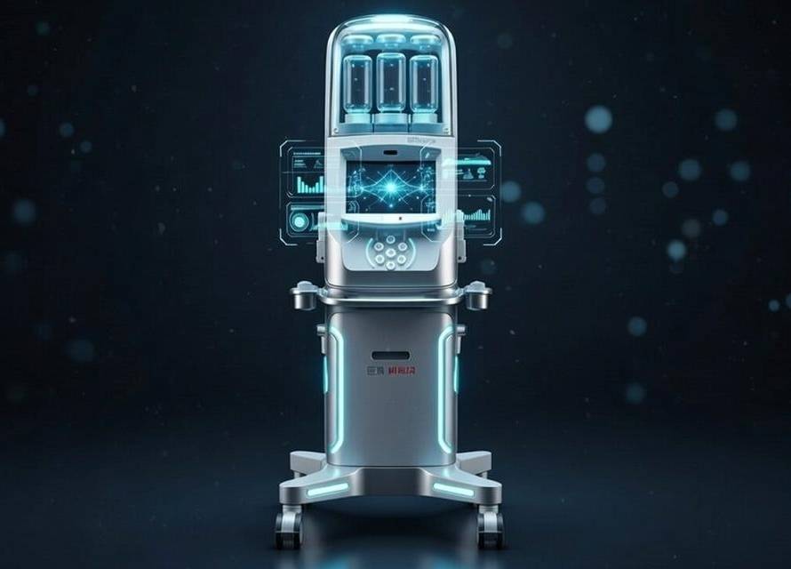
Power injectors deliver 4-5 mL/s iodinated contrast, followed by saline chasers for uniform opacification. Bolus tracking ensures timing; high-concentration agents reduce volume. Dual-head systems like MEDRAD Salient (incorporating SATLine for saline, SATJect for injection) allow precise delivery, minimizing artifacts and venous overlap. Safety protocols address nephrotoxicity and allergies; non-ionic agents preferred. AI predicts optimal injection rates based on patient physiology.
Top 10 Pathologies: Diagnosis, Imaging Protocols, and Tips
| Pathology | Prevalence | Key Features | CTA Protocol | MRA Protocol | Tips and Tricks for Optimal Imaging |
|---|---|---|---|---|---|
| Aneurysms | 2-5% adults | Saccular at CoW junctions; rupture risk 1-2%/year | 120 kV, 0.625 mm slices, MIP reconstructions | TOF 3D, TR 25 ms, flip 20° | CTA: Use volume rendering for neck size; MRA: Avoid flow voids by short TE. CoW variations increase risk—scan entire circle. AI detects growth with 96% sensitivity. |
| Carotid Stenosis | 7-10% >65 | Plaque narrowing >50% | Bolus tracking, curved reformats for NASCET | CE-MRA, dynamic phases | CTA: Dual-energy for plaque type; MRA: Overestimation—correlate with flow. Adjust windows for calcifications. AI automates quantification. |
| Arterial Occlusion | 80% ischemic strokes | Blockage; collaterals via CoW | Arch-to-vertex helical | Phase-contrast for flow | CTA: Source images for thrombus length; MRA: Quantify collaterals. Craniocaudal scan reduces artifacts. AI LVO detection >90% accurate. |
| Arterial Dissection | 2-3/100,000 | Intimal tear, hematoma | Thin slices, multiplanar | Fat-suppressed T1 for hematoma | MRA: High-res for flaps; CTA: Avoid over-injection. Follow-up at 3 months. AI identifies hematomas. |
| Atherosclerosis | Common elderly | Plaque buildup | Dual-energy for vulnerability | Black-blood for wall | CTA: Characterize lipid vs. calcified; MRA: Assess thickening. Use AI for segmentation. |
| Moyamoya Disease | 0.5-1/million | Stenosis with collaterals | Extended coverage for bony | TOF for “puff of smoke” | MRA: Suzuki staging; CTA: Post-op bypass. Respiratory gating reduces artifacts. AI grades collaterals. |
| AVMs | 0.1% | Shunts risking bleed | Dynamic phases | CE for feeders | MRA: Map drainage; CTA: Nidus. Time-resolved for shunting. AI predicts hemorrhage risk. |
| Vasculitis | 15-25/million | Multifocal stenoses | Wall enhancement | High-res for beading | MRA: Monitor therapy; CTA: Biopsy planning. Contrast timing critical. AI detects irregularities. |
| FMD | 0.6-1% females | “String-of-beads” | Confirms morphology | Screens non-invasively | MRA: Young patients—no radiation; CTA: Angioplasty guide. AI classifies patterns. |
| Subclavian Steal | 2-4% extracranial | Flow reversal | Visualizes origin | Phase-contrast reversal | MRA: Duplex complement; CTA: Exercise provocation. CoW collaterals key. AI quantifies reversal. |
For each, CoW assessment is crucial. Tips: Minimize dose, use gating, correlate modalities. AI integration improves detection rates, e.g., CNNs for aneurysm growth surpassing human accuracy (18).
Detailed per pathology: Aneurysms often at ACoA/PCoA junctions; risk factors include hypertension. CTA’s 95% sensitivity aids surgical planning; MRA detects unruptured via TOF. AI models like Rapid Aneurysm offer 96% sensitivity for growth (19). Stenosis: Atherosclerotic; NASCET criteria. MRA overestimates mild cases. AI refines measurements.
Occlusion: Embolic; AI predicts infarct via perfusion maps. Dissection: Traumatic; MRA T1 hyperintensity. Atherosclerosis: Vulnerable plaques; dual-energy CTA. Moyamoya: Genetic; AI stages disease. AVMs: Congenital; AI forecasts rupture. Vasculitis: Autoimmune; AI monitors. FMD: Females; AI screens. Steal: Atherosclerotic; AI flow analysis.
Applications in Clinical Practice
CTA and MRA guide stroke prevention, detecting >50% carotid stenosis for endarterectomy. In aneurysms, CTA identifies saccular types with 90% sensitivity. For dissections, MRA reveals intramural hematomas. Applications extend to screening high-risk populations, with CTA preferred in emergencies. Longitudinal studies show MRA’s utility in monitoring vasculitis response. AI enhances applications by automating triage in telestroke and predicting outcomes.
Case studies: In acute stroke, CTA identifies LVO for thrombectomy; AI platforms like Viz.ai alert teams. For aneurysms, MRA follow-up; AI tracks growth. Guidelines from ACR and AHA endorse multimodal use with AI integration.
Role of SATLine and SATJect in Optimal Image Quality
SATLine and SATJect enable dual-head delivery, reducing venous contamination and artifacts. Supports uniform opacification in CTA, enhancing CoW visualization. Benefits include consistent enhancement; comparisons show 20% artifact reduction.
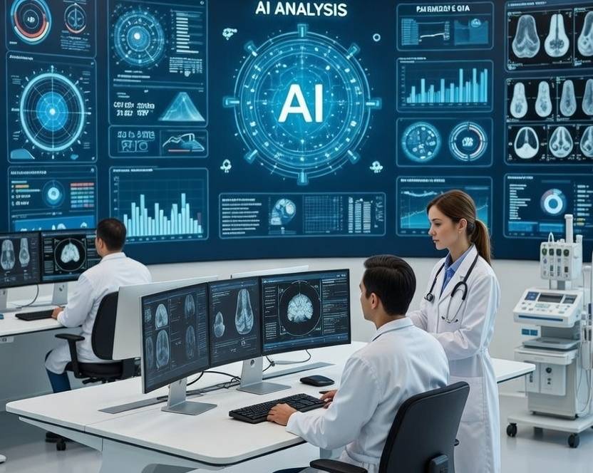
Role of Artificial Intelligence in Cerebrovascular Imaging
AI has transformed vascular imaging by automating detection, segmentation, and prediction in CTA and MRA (13) (16) (17) (18). In stroke imaging, CNNs detect LVOs on CTA with >90% sensitivity, outperforming manual reads in speed (16). Platforms like RapidAI and Viz.ai automate ASPECTS scoring and ICH classification (16). For aneurysms, AI analyzes growth on CTA/MRA, achieving 96% sensitivity vs. 50% for neuroradiologists (19). In MRA, AI refines TOF for CoW variants and collaterals in Moyamoya (17).
Expanded AI Applications in MRA
AI applications in MRA have expanded significantly, particularly in cardiovascular and cerebrovascular contexts. Machine learning optimizes frequency regulation in 3T MRA and corrects artifacts, improving image quality (20). In cancer imaging, AI aids MRA acquisition, reconstruction, registration, and segmentation, enhancing tumor vessel analysis (21). DL accelerates MRA scans, reducing time while maintaining diagnostic accuracy in brain, musculoskeletal, and cardiac applications (22).
For peripheral artery disease (PAD), AI analyzes CE-MRA for microvascular perfusion and skeletal muscle characteristics, achieving 75% accuracy in differentiating PAD patients (23). Non-contrast MRA models leverage AI to avoid gadolinium, reducing risks in renal patients (24). DL shortens MRA scanning via automated positioning and reconstruction, with up to 70% time reduction (25).
Automated coronary MRA systems use AI for exam planning and reconstruction, improving CAD assessment (26). AI impacts MRA pipelines from acquisition to diagnosis, including artifact reduction and prognosis (27). Early AI assesses brain MRA quality and classifies tissues (28).
In clinical MRA, AI detects cerebral aneurysms in TOF-MRA with high sensitivity, retrospectively generates MRA, and synthesizes high-resolution or contrast-weighted images (29). AI advances CMR MRA in planning, acceleration, and post-processing (30). Chatbots interpret MRA reports, classifying tumors and suggesting treatments (31). Overall, AI in MRA enhances efficiency, accuracy, and accessibility, with ongoing developments in non-contrast and perfusion techniques.
Applications include infarct segmentation, outcome prediction (e.g., hemorrhagic transformation), and collateral grading (17). Challenges: Data variability, overfitting, ethical issues like bias. Future: Transformers and LLMs for multimodal analysis, XR-robotic integration (13). Evidence from 2023-2026 reviews shows AI’s preclinical promise, with RCTs supporting recovery-phase tools (16).
AI architectures: CNNs for object detection; U-Net for segmentation. In AIS, AI estimates onset via DWI-FLAIR mismatch (AUC 0.814) (18). For IAs, PointNet++ predicts rupture. AVMs: Random forest for risk. Translational hurdles: FDA approval, integration into PACS.
Multimodal integration improves outcomes; AI for variants. Limitations: CTA radiation, MRA cost. Geographic trends: Higher variants in Africans. Controversies: AI bias in diverse populations; ethical data use. Future: AI-robotic convergence for telerobotics.
Conclusion
CTA and MRA are indispensable for CoW and carotid imaging, with optimized protocols and AI enhancing diagnostics. AI integration promises precision, but requires addressing challenges for widespread adoption.
References
- Cleveland Clinic. (2024). Circle of Willis. https://my.clevelandclinic.org/health/body/circle-of-willis
- Klimek-Piotrowska, W., et al. (2016). A multitude of variations in the configuration of the circle of Willis. Anatomical Science International, 91(4), 325-333.
- Josephson, S. A., et al. (2004). Evaluation of carotid stenosis using CT angiography. American Journal of Neuroradiology, 25(7), 1197-1202.
- Medical News Today. (2020). Circle of Willis: Anatomy, function, and what to know. https://www.medicalnewstoday.com/articles/circle-of-willis
- Radiopaedia. (2018). CT angiography of the cerebral arteries (protocol). https://radiopaedia.org/articles/ct-angiography-of-the-cerebral-arteries-protocol?lang=us
- Stroke Manual. (2025). CT angiography (CTA). https://www.stroke-manual.com/ct-angiography-cta
- PMC. (2020). Examination of Structural Variations of the Circle of Willis by 3D-TOF-MRA. https://pmc.ncbi.nlm.nih.gov/articles/PMC7026468
- PMC. (2021). Contrast-Enhanced MR Angiography of the Carotid and Vertebrobasilar Systems. https://pmc.ncbi.nlm.nih.gov/articles/PMC8148843
- Journal of the American College of Radiology. (2017). ACR Appropriateness Criteria® Cerebrovascular Disease. https://www.jacr.org/article/S1546-1440(17)30210-7/fulltext
- Endovascular Today. (2009). CTA and the Circle of Willis. https://evtoday.com/articles/2009-may/EVT0509_03-php
- Nature. (2020). Quantitative Analysis of the Cerebral Vasculature on Magnetic Resonance Angiography. https://www.nature.com/articles/s41598-020-67225-w
- PMC. (2013). A Comprehensive Study of the Anatomical Variations of the Circle of Willis. https://pmc.ncbi.nlm.nih.gov/articles/PMC3879841
- Sorkin, G. C., et al. (2026). Current state of the clinical applications of artificial intelligence in stroke: A literature review. Brain Sciences, 16(2), 173. https://doi.org/10.3390/brainsci16020173
- Trando Med. (2023). What diseases are associated with variations in the Circle of Willis?https://www.trando-med.com/info/what-diseases-are-associated-with-variations-i-85057759.html
- Stroke Manual. (2025). Anatomical variants of cerebral arteries. https://www.stroke-manual.com/anatomical-variants-of-cerebral-arteries
- Liu, Y., et al. (2024). Artificial intelligence in ischemic stroke images: Current applications and future directions. Frontiers in Neurology, 15. https://doi.org/10.3389/fneur.2024.1418060
- Sen, R. D., & Levitt, M. R. (2024). Artificial intelligence in neurointerventions. Endovascular Today. https://evtoday.com/articles/2024-june/artificial-intelligence-in-neurointerventions
- ATrainCEU. (n.d.). 4. Carotid and Vertebrobasilar Disorders. https://www.atrainceu.com/content/4-carotid-and-vertebrobasilar-disorders
- Hall, J. (2026, February 6). Is AI better than neuroradiologists at evaluating aneurysm growth on CTA and MRA scans? Diagnostic Imaging. https://www.diagnosticimaging.com/view/ai-neuroradiologists-evaluating-aneurysm-growth-cta-mra-scans-
- The Applications of Artificial Intelligence in Cardiovascular Magnetic Resonance—A Comprehensive Review. (2022). PMC. https://pmc.ncbi.nlm.nih.gov/articles/PMC9147423
- A critical assessment of artificial intelligence in magnetic resonance imaging of cancer. (2025). Nature. https://www.nature.com/articles/s44303-025-00076-0
- Artificial intelligence in MRI clinical practice: From historical innovation to emerging trends. (n.d.). ScienceDirect. https://www.sciencedirect.com/science/article/pii/S1687850726000014
- Innovations in MRI and AI Integration for Vascular Plaque Evaluation and Overview of Deep Learning Techniques in Peripheral Vascular Disease. (n.d.). Methodist DeBakey Cardiovascular J. https://journal.houstonmethodist.org/articles/10.14797/mdcvj.1642
- Artificial Intelligence Applications in Cardiovascular Magnetic Resonance Imaging: Are We on the Path to Avoiding the Administration of Contrast Media? (n.d.). MDPI. https://www.mdpi.com/2075-4418/13/12/2061
- Deep learning and AI in reducing magnetic resonance imaging scanning time: advantages and pitfalls in clinical practice. (n.d.). Polish Journal of Radiology. https://www.polradiol.com/Deep-learning-and-AI-in-reducing-magnetic-resonance-imaging-scanning-time-advantages,192822,0,2.html
- AI Enhances Coronary MR Angiography for CAD Assessment. (2025). Journal of Cardiovascular Magnetic Resonance. https://www.linkedin.com/posts/journal-of-cardiovascular-magnetic-resonance-892240191_whycmr-activity-7405000042149015553-TmCR
- The intelligent imaging revolution: artificial intelligence in MRI and MRS acquisition and reconstruction. (2024). PMC. https://pmc.ncbi.nlm.nih.gov/articles/PMC11316718
- Deep Learning Artificial Intelligence (AI) Applications in MRI. (2019). McGovern Medical School. https://med.uth.edu/radiology/2019/10/02/deep-learning-artificial-intelligence-ai-applications-in-mri
- Advancing clinical MRI exams with artificial intelligence: Japan’s contributions and future prospects. (2024). Japanese Journal of Radiology. https://link.springer.com/article/10.1007/s11604-024-01689-y
- Improving the efficiency and accuracy of cardiovascular magnetic resonance with artificial intelligence—review of evidence and proposition of a roadmap to clinical translation. (n.d.). ScienceDirect. https://www.sciencedirect.com/science/article/pii/S1097664724010780
- Application of artificial intelligence chatbots in interpreting magnetic resonance imaging reports: a comparative study. (2025). Nature. https://www.nature.com/articles/s41598-025-17355-w