Introduction
Cardiac CT angiography (CCTA), also known as coronary CT angiography or heart CT scan for blocked arteries, is the leading non-invasive imaging test for coronary artery disease (CAD) and coronary stenosis evaluation in 2026. This comprehensive guide combines the latest advancements in CCTA diagnostic accuracy (including positive predictive value (PPV) and negative predictive value (NPV) by stenosis severity), optimal contrast flow rates and injection protocols, CT-FFR for hemodynamic significance, AI-powered plaque morphology analysis and outcome prediction, and the latest SCCT guidelines (CAD-RADS 2.0 with recent quantitative and AI updates) [1,2].
Whether you’re researching CCTA vs invasive coronary angiography, how CCTA rules out blocked arteries, or AI in coronary plaque assessment, this guide provides everything you need—updated for February 2026 with current trends, reimbursement changes, and clinical insights.
What Is Cardiac CT Angiography (CCTA)?
Cardiac CT angiography (CCTA) is a non-invasive imaging procedure that uses iodinated contrast and computed tomography to visualize the coronary arteries with high resolution. It accurately detects coronary stenosis, plaque buildup, calcification, and high-risk features without catheterization. As a first-line test for patients with intermediate pretest probability of CAD or chest pain, CCTA offers lower radiation exposure, rapid results, and excellent rule-out capability compared to invasive coronary angiography [3].
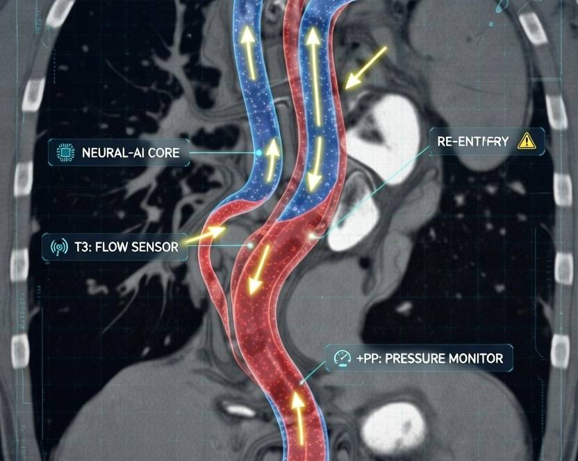
Optimal Contrast Flow Rates and Injection Protocols in CCTA 2026
Diagnostic-quality CCTA requires homogeneous coronary opacification (>300-350 HU). Modern protocols use high-concentration iodine contrast (320-400 mgI/mL) with power injection and saline chasers [3,4].
| Protocol Type | Flow Rate (mL/s) | Contrast Volume (mL) | Saline Chaser (mL) | Key Benefits & Clinical Notes |
|---|---|---|---|---|
| Standard Monophasic | 5-6 | 50-80 | 30-50 (same rate) | Reliable enhancement; widely used in practice [3] |
| Biphasic/Triphasic | 4-7 | 60-100 | 20-40 | Reduces venous artifacts; patient-tailored |
| High-Flow for Advanced Scanners | 5-7 | 50-70 | 30-40 | Superior for obese patients or low-kVp protocols [4] |
| Personalized (Weight/kVp-Based) | 4-6.5 | 0.8-1.2 mL/kg | Matched rate | Optimizes attenuation; minimizes contrast volume |
High flow rates (≥5 mL/s) create a compact bolus for sharp vessel delineation and fewer artifacts. SCCT guidelines recommend 5-7 mL/s with bolus tracking for optimal coronary visualization [3].
CCTA Diagnostic Accuracy: PPV and NPV by Coronary Stenosis Severity
CCTA demonstrates outstanding performance versus invasive angiography, with sensitivity 95-98%, specificity 80-90%, and NPV 95-99% in recent meta-analyses [5,6].
| Stenosis Severity | Sensitivity (%) | Specificity (%) | PPV (%) | NPV (%) | Key Clinical Insights |
|---|---|---|---|---|---|
| ≥50% (Obstructive) | 95-97 | 83-90 | 64-88 | 95-99 | High NPV safely excludes obstructive CAD [5] |
| 50-69% (Intermediate) | 85-92 | 85-92 | 50-70 | >95 | Frequent overestimation; functional testing (CT-FFR) recommended [6] |
| ≥70% (Severe) | 93-98 | 90-95 | 80-92 | 97-99 | Strong PPV; high likelihood of ischemia |
| ≥90% (Critical) | >95 | >95 | >90 | >98 | Highly predictive of hemodynamically significant lesions |
PPV increases with stenosis severity due to fewer false positives in high-grade lesions, while NPV remains consistently excellent—making CCTA an ideal gatekeeper to invasive procedures [5].
CT-Derived Fractional Flow Reserve (CT-FFR): Functional Assessment in 2026
CT-FFR computes fractional flow reserve from standard CCTA datasets using AI or computational fluid dynamics, identifying ischemia-causing lesions (≤0.80 significant) without additional scanning or stress [7,8].
| Metric vs. Invasive FFR | Value (%) | Clinical Advantages |
|---|---|---|
| Sensitivity | 85-92 | Excellent detection of functional ischemia [7] |
| Specificity | 75-90 | Superior to anatomic CCTA, especially intermediate stenoses |
| Overall Accuracy | 80-87 | Reclassifies 30-50% of positive CCTA findings |
| Correlation | r=0.80-0.90 | Strongest agreement in 70-90% stenosis |
CT-FFR significantly improves specificity for intermediate lesions, reduces unnecessary invasive angiography, and aligns with ACC/AHA and SCCT guidelines [7].
Artificial Intelligence (AI) in CCTA: Plaque Morphology Analysis and Prognostic Outcomes
AI quantitative plaque analysis automates stenosis grading, plaque segmentation, and identification of high-risk features (e.g., low-attenuation plaque, necrotic core) on CCTA [9,10].
| AI Application | Performance | Prognostic Impact & 2026 Trends |
|---|---|---|
| Stenosis Detection/Quantification | AUC 0.93-0.96 | Faster, expert-level accuracy [9] |
| Plaque Volume & Composition | r>0.90 vs. IVUS | Precise non-calcified/low-attenuation measurement |
| High-Risk Plaque Features | Sensitivity >90% | Detects vulnerable plaques [10] |
| MACE Prediction | HR 1.5-3.0 | Incremental risk beyond stenosis; guides intensive prevention |
In 2026, AI plaque analysis earns improved reimbursement (Category I codes), enabling broader adoption for long-term risk stratification independent of stenosis severity [9].
Latest SCCT Guidelines 2026: CAD-RADS 2.0 and Recent Updates
CAD-RADS 2.0 (2022) standardizes CCTA reporting with stenosis severity, plaque burden, and modifiers (HRP for high-risk plaque, I for ischemia via CT-FFR) [2].
| CAD-RADS Category | Stenosis Severity | Management Recommendation |
|---|---|---|
| 0 | None | No further evaluation |
| 1-2 | 1-49% | Guideline-directed medical therapy |
| 3 | 50-69% | Consider functional testing or CT-FFR |
| 4A/4B | 70-99% / LM >50% | ICA consideration |
| 5 | Total occlusion | ICA recommended |
| 6 | Non-diagnostic | Alternative testing |
Recent updates include quantitative plaque standards and AI integration emphasis [2,11].
CCTA vs Invasive Coronary Angiography
CCTA provides comparable or superior outcomes with lower procedural risks, reduced downstream revascularization, and non-invasive advantages. Ideal for stable chest pain evaluation; invasive angiography reserved for high-risk cases or intervention [12].
Clinical Implications of CCTA in 2026
With optimized high-flow protocols, CCTA reliably excludes obstructive CAD (high NPV), while severe stenosis or positive CT-FFR yields strong PPV for intervention. AI plaque analysis adds powerful prognostic value, identifying high-risk patients early. Following SCCT guidelines minimizes invasive procedures and personalizes CAD management [1,2,7].
FAQs: Cardiac CT Angiography (CCTA) 2026
- What is the difference between cardiac CT and CCTA? Standard cardiac CT assesses structure/function; CCTA specifically images coronary arteries with contrast for stenosis/plaque evaluation.
- Is CCTA better than invasive coronary angiography? Yes for diagnosis and risk stratification—non-invasive, lower risk, high NPV, and excellent outcomes.
- How accurate is CCTA for detecting blocked arteries? NPV >95-99%; outstanding rule-out capability [5].
- What is CT-FFR and does insurance cover it? Non-invasive functional assessment from CCTA data; widely reimbursed [7].
- Is AI plaque analysis covered by insurance in 2026? Yes, with improving Category I reimbursement [9].
- Who should get a CCTA? Patients with intermediate-risk chest pain or suspected CAD—consult your cardiologist.
- Updated February 2026 with the latest SCCT guidelines, AI trends, and clinical evidence.
References
- Abbara, S., Blanke, P., Cury, R. C., Leipsic, J., Maroules, C., Rossi, A., … & SCCT Guidelines Committee. (2016). SCCT guidelines for the performance and acquisition of coronary computed tomographic angiography: A report of the Society of Cardiovascular Computed Tomography Guidelines Committee. Journal of Cardiovascular Computed Tomography, 10(6), 435-449. https://doi.org/10.1016/j.jcct.2016.10.002
- Cury, R. C., Abbara, S., Achenbach, S., Agatston, A., Berman, D. S., Budoff, M. J., … & Shaw, L. J. (2022). CAD-RADS™ 2.0 – 2022 Coronary Artery Disease – Reporting and Data System: An expert consensus document of the Society of Cardiovascular Computed Tomography (SCCT). Journal of Cardiovascular Computed Tomography, 16(4), 361-381. https://doi.org/10.1016/j.jcct.2022.07.002
- Abbara et al. (2016). (as reference 1)
- Bae, K. T. (2010). Intravenous contrast medium administration and scan timing at CT: Considerations and approaches. Radiology, 256(1), 32-61. https://doi.org/10.1148/radiol.10090908
- Meta-analysis of the diagnostic accuracy of computed tomography angiography compared with invasive coronary angiography. (2025). Various sources including PMC.
- Celeng, C., et al. (2019). Anatomical and functional computed tomography for diagnosing coronary artery disease. European Heart Journal – Cardiovascular Imaging.
- Tang, C. X., et al. (2024). A meta-analysis comparing the diagnostic performance of computed tomography fractional flow reserve and coronary computed tomography angiography. European Radiology.
- Nørgaard, B. L., et al. (2022). Diagnostic performance of noninvasive fractional flow reserve derived from coronary computed tomography angiography. Various JACC.
- Narula, J., et al. (2025). Quantitative coronary plaque analysis in clinical practice. JACC: Cardiovascular Imaging.
- Choi, A. D., et al. (2025). Artificial intelligence–based coronary plaque quantification. JACC: Cardiovascular Imaging.
- Society of Cardiovascular Computed Tomography. (2023-2025 updates). Quantitative standards and AI integration.
- Knuuti, J., et al. (2019). 2019 ESC Guidelines for the diagnosis and management of chronic coronary syndromes. European Heart Journal.